Info
devi rubare gli appunti dall’ari e viola.
TARGET DECK: MED::I::Signaling Pathways in Health and Disease::Cell Signaling::02 - Cell surface receptors and nuclear receptors
Overview
Lecture Topics
- Types of ligands & receptors
- Principles of ligand-receptor interactions
- Pathways of cellular signaling
- Molecular details of representative signal transduction systems, classified by receptor type
Classification of Receptors
Membrane Receptors
- Ionotropic receptors (ion channels)
- Metabotropic receptors (G proteins)
- Catalytic receptors (enzymes)
Intracellular Receptors
- Nuclear receptors (e.g., steroid receptors)
Ionotropic Receptors — Overview
Definition
Ionotropic receptors are the simplest transducers of extracellular signals (ligands). They are ligand-gated ion channels that directly open or close in response to the concentration of a signal ligand or membrane potential.
Examples:
- Nicotinic acetylcholine receptor (nAChR)
- receptor
- Glycine receptor
- Glutamate receptor
- Serotonin receptor
What are ionotropic receptors?
Ligand-gated ion channels that directly convert chemical signals (ligand binding) into electrical/ionic changes. They are the simplest transducers of extracellular signals.
Anki cloze
Ionotropic receptors are also called {1:ligand-gated ion channels} and are considered the {2:simplest} transducers of extracellular signals.
Acetylcholine at the Neuromuscular Junction
The Cholinergic Synapse — Step by Step
- An action potential travels down the presynaptic axon, triggering opening of voltage-gated channels
- influx causes secretory vesicles containing acetylcholine to fuse and release ACh into the synaptic cleft
- ACh diffuses across the cleft and binds to nicotinic receptors on the postsynaptic membrane (myocyte or next neuron)
- The ligand-gated cationic channel opens (~1 ms), allowing extracellular and to enter
- Ion influx depolarizes the postsynaptic cell (changes ), triggering muscle contraction
Role of Ions as Second Messengers
Extracellular and entering through the nicotinic channel act as intracellular second messengers, depolarizing the postsynaptic cell and affecting other membrane proteins sensitive to .
CC(=O)OCC[N+](C)(C)C(Acetylcholine)
What is the role of acetylcholine at the neuromuscular junction?
ACh is a key excitatory neurotransmitter that binds to nicotinic receptors (ligand-gated cationic channels) on the postsynaptic membrane, causing channel opening, Na⁺/Ca²⁺ influx, membrane depolarization, and ultimately muscle contraction.
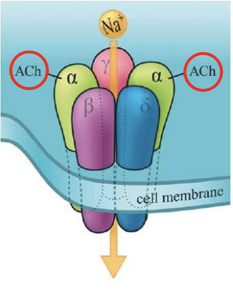
Nicotinic Acetylcholine Receptor (nAChR)
Found in the post-synaptic surface. The pre-synaptic neuron will produce and release acetylcholine in the cleft between the synapses (extracellular space). They will find a binding site in the post-synaptic cell, advising it to continue the link.
Acetylcholine is not an ion: it does not go through. It opens the channel for positively charged ions, sodium and calcium (specificity).
The channel is a multi-subunit protein, pentameric → .
The alpha subunits get their conformation changed by the binding of acetylcholine, inducing the other subunits to become “larger”.
The gets rotated.
Localization
- Neuromuscular junctions
- Sympathetic and parasympathetic ganglia
- Adrenal medulla
- Central nervous system
Structure
Molecular Architecture
- Pentameric structure: subunits
- Each subunit contains four transmembrane -helices (M1–M4)
- The M2 helix of each subunit lines the ion-conducting pore
| Feature | Detail |
|---|---|
| Subunit composition | (pentamer) |
| Transmembrane helices per subunit | 4 (M1–M4) |
| Pore-lining helix | M2 |
| Ion selectivity | Cations (, , ) |
| Ligand binding site | Two ACh molecules bind on the -subunits |
Key Reference
Miyazawa A, Fujiyoshi Y, Unwin N. Structure and gating mechanism of the acetylcholine receptor pore. Nature. 2003, 423: 949–955.
What is the subunit composition of the nicotinic acetylcholine receptor?
The nAChR is a pentamer composed of α₂βγδ subunits, each containing four transmembrane α-helices (M1–M4). The M2 helix of each subunit lines the pore.
Anki cloze
The nicotinic ACh receptor has a {1:pentameric} structure composed of {2:α₂βγδ} subunits, each with {3:four} transmembrane α-helices. The pore is lined by the {4:M2} helices.
Channel Gating Mechanism
Structural Basis of Gating
In the closed (resting) state: the M2 helices are oriented such that the side chains of five Leucine residues (one per subunit, at position Leu-257 on M2, i.e. α-Leu257) form a hydrophobic ring at the middle of the membrane, physically occluding the channel pore.
Upon binding of 2 ACh molecules to the -subunits, an allosteric conformational change is induced: the M2 helices rotate, moving the Leu side chains away from the pore center and exposing polar residues, thereby opening the passage to cations.
What is the structural basis of nAChR channel gating?
In the resting state, five Leu residues (one per M2 helix, α-Leu257) form a hydrophobic ring that closes the pore. ACh binding induces an allosteric rotation of the M2 helices, moving the Leu side chains away from the center and exposing polar residues, thus opening the channel to cations.
Functional States of nAChR
| State | ACh Binding | Pore | Conductance |
|---|---|---|---|
| Resting | No | Closed | Non-conducting |
| Activated | Yes (2 ACh on α-subunits) | Open | Conducting (cations) |
| Desensitized | Yes (usually occupied) | Closed | Non-conducting |
Desensitization
The acetylcholine signal is transient: the receptor possesses an intrinsic mechanism that closes the channel even in the continued presence of ACh. In the desensitized state, the ACh binding sites are typically occupied but the pore is closed and non-conducting.
What is desensitization of the nAChR?
Desensitization is a state in which the receptor has ACh bound but the channel is closed and non-conducting. It acts as an intrinsic timer that limits the duration of the signal even in the continued presence of the agonist.
Anki cloze
In the {1:desensitized} state of the nAChR, the ACh binding sites are {2:occupied} but the pore is {3:closed} and non-conducting.
Signal Termination — Acetylcholinesterase
Enzymatic Hydrolysis of ACh
Acetylcholinesterase (AChE) catalyzes the hydrolysis of ACh into choline and acetate, allowing the cholinergic neuron to return to its resting state after activation.
- Choline is recycled back into the nerve terminal via a choline carrier
- This ensures rapid termination of the cholinergic signal
Reference
Dvir H, Silman I, Harel M, Rosenberry TL, Sussman JL. (2010) Chem Biol Interact. 187:10–22.
What enzyme terminates the ACh signal at the synapse, and what are its products?
Acetylcholinesterase (AChE) hydrolyzes ACh into choline and acetate. Choline is recycled back into the presynaptic neuron via a carrier.
Anki cloze
Acetylcholinesterase hydrolyzes acetylcholine into {1:choline} and {2:acetate}, allowing the cholinergic neuron to return to its {3:resting} state.
Receptor
Overview
The receptor is an ionotropic receptor gated by GABA (-aminobutyric acid). It is a ligand-gated chloride channel; influx hyperpolarizes the membrane (inhibitory effect), in contrast to the depolarizing cation channels of nAChR.
Key Concept: Depolarization vs. Hyperpolarization
Important
Ligands that stimulate ion channels can either depolarize or hyperpolarize the target cell membrane, depending on the type of ion that passes through the pore:
- / influx → Depolarization (excitatory)
- influx → Hyperpolarization (inhibitory)
Anki cloze
The GABA_A receptor, upon activation, allows {1:Cl⁻} to flow into the cell, causing {2:hyperpolarization} of the membrane, which is an {3:inhibitory} effect.
Pharmacological Modulation of
Allosteric Modulation Sites
Several pharmacological substances can modulate GABA’s effect by binding to sites distinct from the GABA binding site on the receptor.
| Substance Class | Effect on GABA Activity | Functional Outcome |
|---|---|---|
| Benzodiazepines | Increase | Anxiolytic |
| Barbiturates | Increase | Sedative/anesthetic |
| Ethanol | Increase | CNS depression |
| Pregnenolone sulfate / Allopregnanolone / Allotetrahydrodeoxycorticosterone (neuroactive steroids) | Increase | Sedative |
| -carbolines | Decrease | Anxiogenic |
| Picrotoxin / TBPS | Decrease (channel block) | Convulsant |
| Bicuculline | Decrease (competitive antagonist) | Convulsant |
| Flumazenil | Blocks benzodiazepine site | Reversal agent |
| Ciclopirroloni / Imidazopiridine | Increase (BZD site agonists) | Anxiolytic/hypnotic |
Receptor
Regulation receptor activity is also regulated by phosphorylation by PKA and PKC.
Name two classes of substances that increase GABA_A activity and two that decrease it.
Increase: Benzodiazepines, barbiturates (also ethanol, neuroactive steroids).
Decrease: β-carbolines, picrotoxin/bicuculline.
Anki cloze
Flumazenil acts at the {1:benzodiazepine} binding site of GABA_A and is used to {2:reverse} the effects of benzodiazepines. β-carbolines are {3:inverse agonists} that reduce GABA_A activity and are {4:anxiogenic}.
Comparison of Major Receptor Types
| Receptor Type | Mechanism | Second Messenger | Example |
|---|---|---|---|
| Ionotropic (ion channel) | Direct ion flow on ligand binding | Ions (, , ) | nAChR, |
| Metabotropic (GPCR) | G protein → enzyme → second messenger | cAMP, , DAG | mAChR, -adrenergic |
| Catalytic (receptor enzyme) | Ligand binding activates intracellular enzyme domain | Phosphorylation cascades | Receptor tyrosine kinases |
| Receptor with no intrinsic enzyme | Interacts with cytosolic kinase → gene regulation | Kinase cascades | Cytokine receptors |
| Nuclear/intracellular | Steroid binds nuclear receptor → regulates gene expression | mRNA / protein | Steroid receptors |
Nuclear / Intracellular Receptors
Mechanism
Steroid hormones (lipophilic) diffuse through the plasma membrane and bind to nuclear receptor proteins. The ligand-receptor complex then acts as a transcription factor, regulating the expression of specific target genes → production of specific mRNA and proteins.
How do nuclear receptors differ from membrane receptors in their mechanism of action?
Nuclear receptors bind lipophilic ligands (e.g., steroids) intracellularly. The ligand-receptor complex directly regulates gene expression by acting as a transcription factor, in contrast to membrane receptors which transduce signals via second messengers or kinase cascades.
Mnemonic — Ionotropic Receptor Ligands:
“A GABA eats Gly and 5-HT”
- Acetylcholine (nicotinic)
- GABA ()
- Glutamate
- Glycine
- 5-HT (Serotonin)
TLDR
Lecture A.02 — Key Points
- Receptors are classified as membrane (ionotropic, metabotropic, catalytic) or intracellular (nuclear)
- Ionotropic receptors are ligand-gated ion channels — the simplest signal transducers; they directly convert ligand binding into ion flux and membrane potential changes
- nAChR is a pentameric () cationic channel; ACh binding causes allosteric rotation of M2 helices (moving Leu257 away from pore center) → channel opens; found at NMJ, ganglia, adrenal medulla, CNS
- nAChR states: resting (unoccupied, closed) → activated (ACh bound, open) → desensitized (ACh bound, closed) — desensitization terminates the signal intrinsically
- Acetylcholinesterase hydrolyzes ACh → choline + acetate, enabling signal termination; choline is recycled
- is an inhibitory ionotropic receptor: influx → hyperpolarization; can be allosterically modulated by benzodiazepines (↑), barbiturates (↑), ethanol (↑), neuroactive steroids (↑), -carbolines (↓), picrotoxin/bicuculline (↓); regulated by PKA/PKC phosphorylation
- Ion type determines effect: cation influx (/) → depolarization (excitatory); influx → hyperpolarization (inhibitory)
- Nuclear receptors bind lipophilic ligands (steroids), translocate to nucleus, and directly regulate gene transcription