1. Glandular Epithelia — Overview
A gland is one or more cells that produce and secrete a specific product. The product is always a water-based (aqueous) fluid, usually containing proteins — referred to as a secretion.
Classification by Secretion Destination
| Type | Description |
|---|---|
| Endocrine | Ductless; secretes into blood or lymphatic vessels |
| Exocrine | Secretes via ducts onto an epithelial surface (skin or body cavity) |
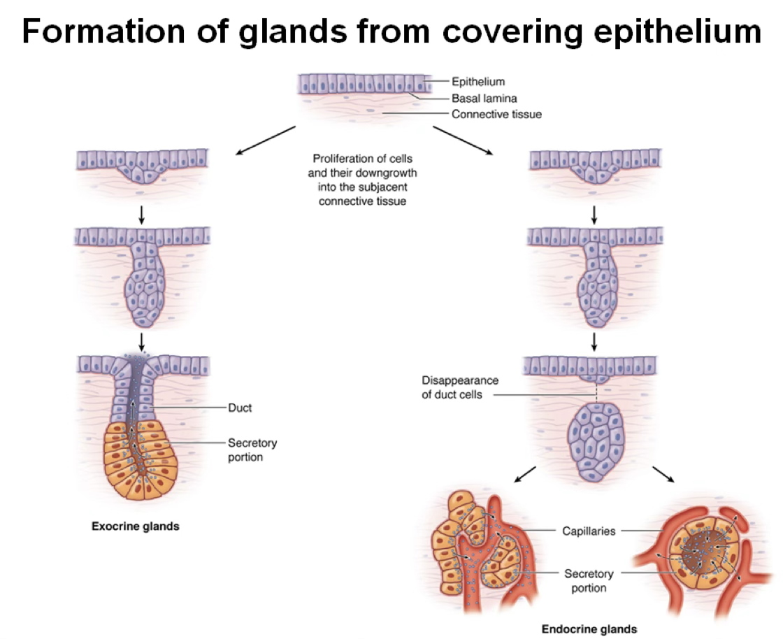
Gland Genesis
Glands form when epithelial cells proliferate and invade the underlying connective tissue migrating through the basement. After the migration is completed they will start do differentiate into the parenchima of the gland (secretory portion).
- Exocrine glands will keep the connection to the surface epithelium, representing the duct.
- Endocrine glands lose their ductal connection to the originating epithelium.
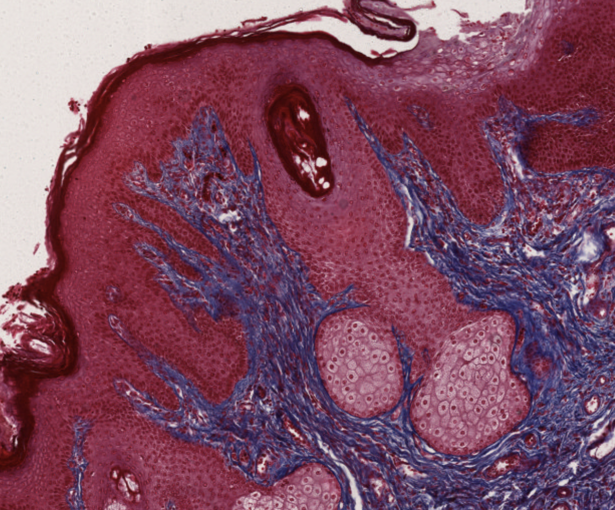
Classify this Gland
unibo.smartzoom.com
This is squamous pluristratified epithelium. There are glands, both exocrine, with the ducts, and endocrine. The ducts are not yet mature, since we would se a columnar/cuboidal epithelium.
2. Exocrine Glands — Structure
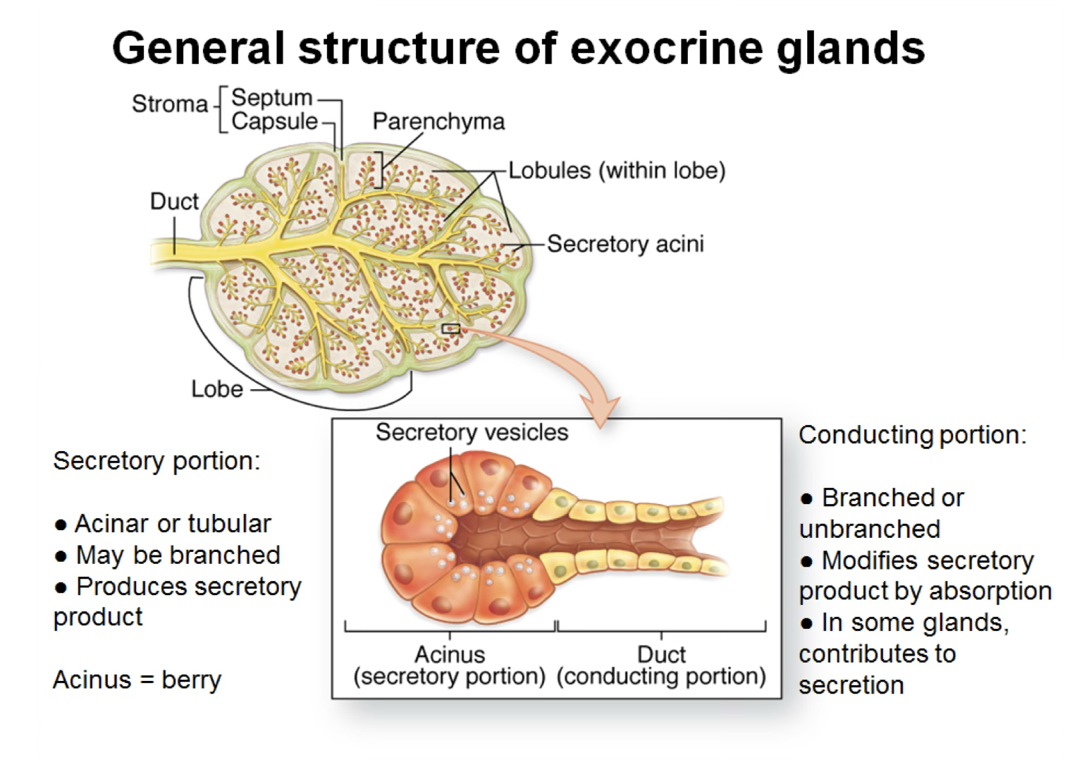
- Large multicellular glands are surrounded by a collagenous connective tissue capsule
- The capsule sends septa into the gland, subdividing it into lobes and lobules
Secretory Granules
Glandular epithelia manufacture products by synthesizing macromolecules, which are packaged and stored in secretory granules. Products include:
- Polypeptide hormones (e.g., EGF)
- Waxy substances
- Mucinogen (goblet cells)
- Milk (lipids + proteins + carbohydrates) — mammary glands
- Cytokines for cell–cell communication
3. Types of Signaling (by Distance)
| Mode | Description |
|---|---|
| Autocrine | Ligand acts on its own producer cell |
| Paracrine | Target cell is in the vicinity of the signaling cell |
| Endocrine | Target cell is distant; cytokine travels via blood/lymph |
4. Classification of Exocrine Glands
Exocrine glands are classified by:
- Number of secreting cells
- Unicellular Glands (like Goblet) → Multicellular Glands
- Nature of secretion
- Mode of secretion
5. Unicellular Glands — Goblet Cell
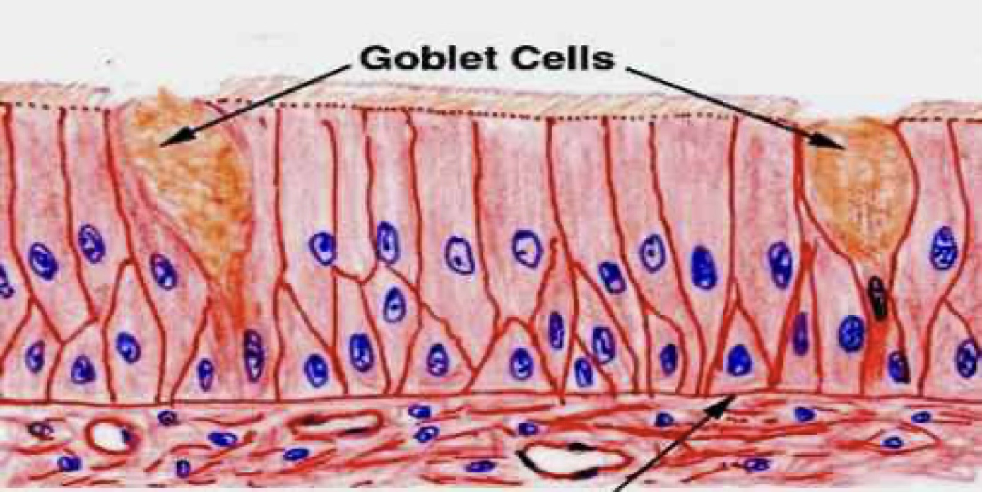
Goblet Cell Structure
- Thin basal region sits on the basal lamina
- Expanded apical portion (theca) faces the lumen of the digestive tube or respiratory tract
- The theca is filled with secretory droplets of mucinogen, displacing the cytoplasm to the periphery and the nucleus to the base
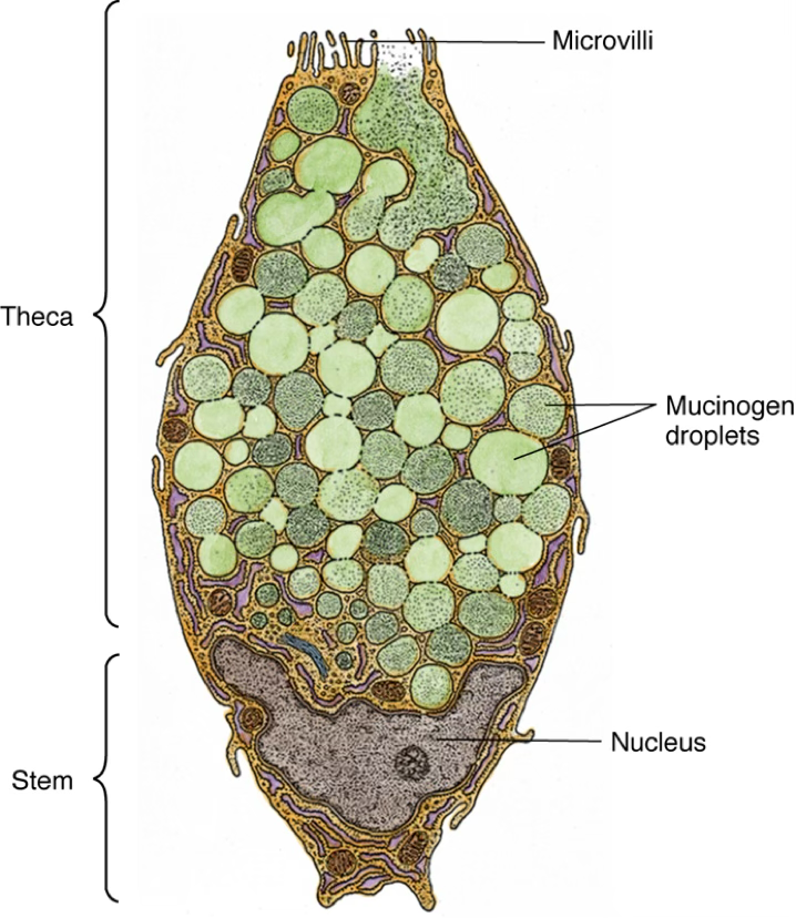
- Release is regulated by chemical irritation and parasympathetic innervation
- Results in exocytosis of the entire secretory contents → protects the epithelial sheet
Classifying Goblet
https://unibo.smartzoom.com/s1241/course1776/f1791/i1794/
Nucleus always found at the bottom. You can also see goblet cells that didn’t reach the surface.
The blue droplets are the goblet cells.
6. Classification by Morphology
6.1 Simple vs. Compound
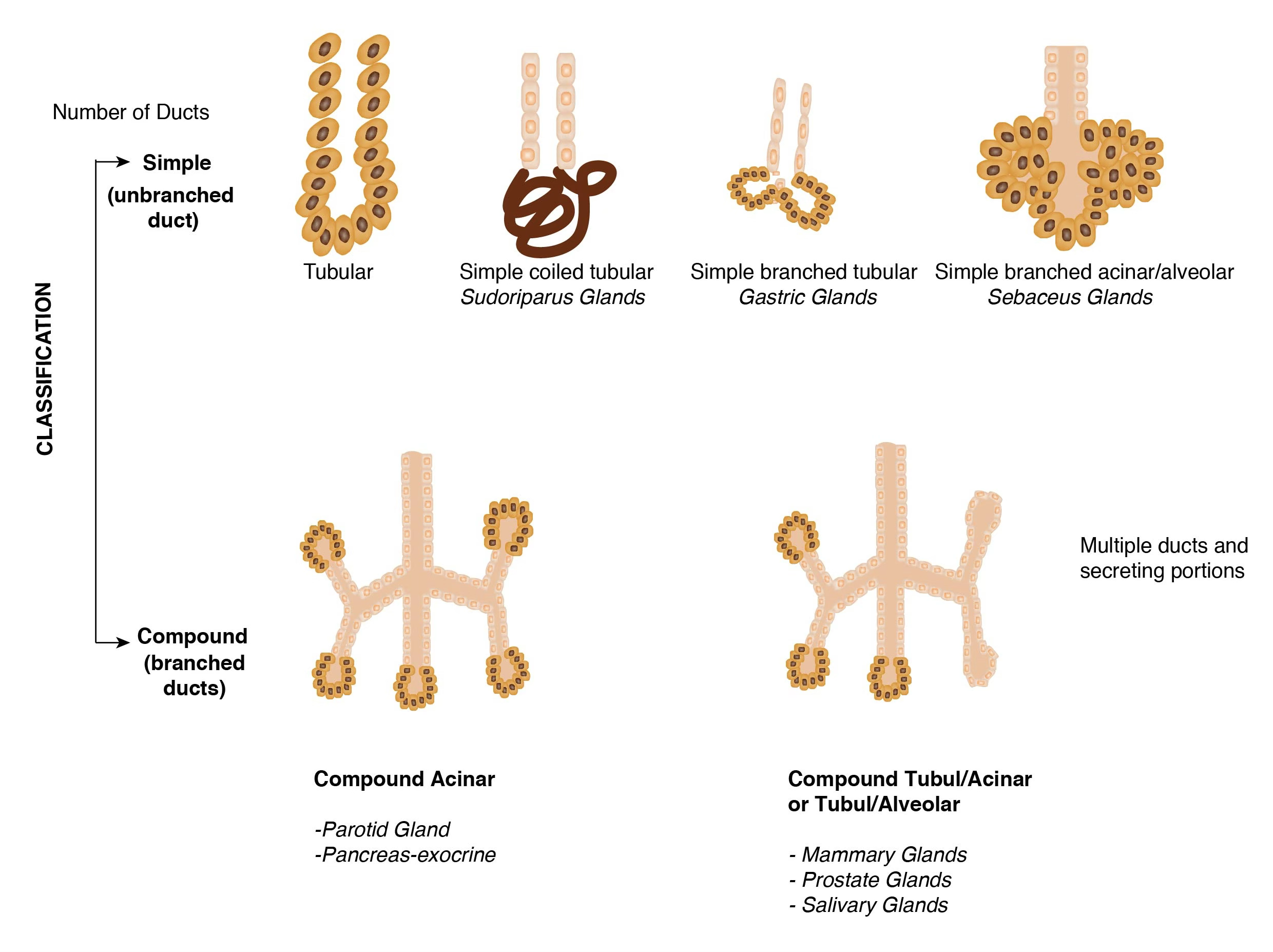
| Category | Description |
|---|---|
| Simple | Unbranched duct |
| Compound | Branched duct system |
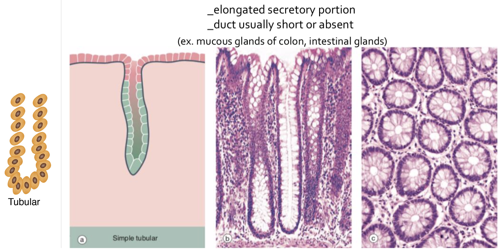
6.2 Shape of Secretory Portion
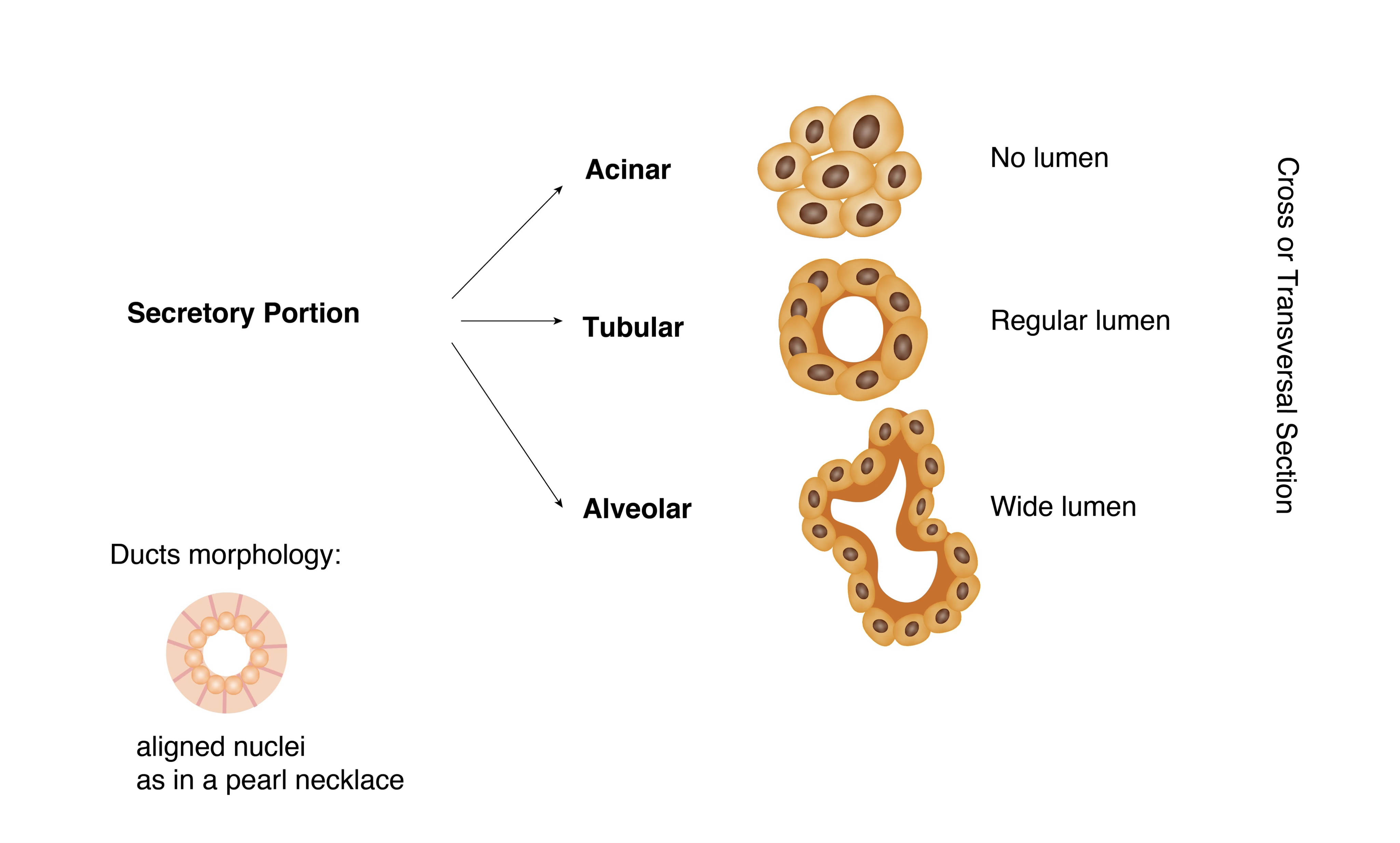
| Shape | Description |
|---|---|
| Tubular | Tube-shaped secretory unit |
| Acinar / Alveolar | Flask- or sac-shaped secretory unit |
| Tubulo-acinar | Mixed |
7. Type of Secretion
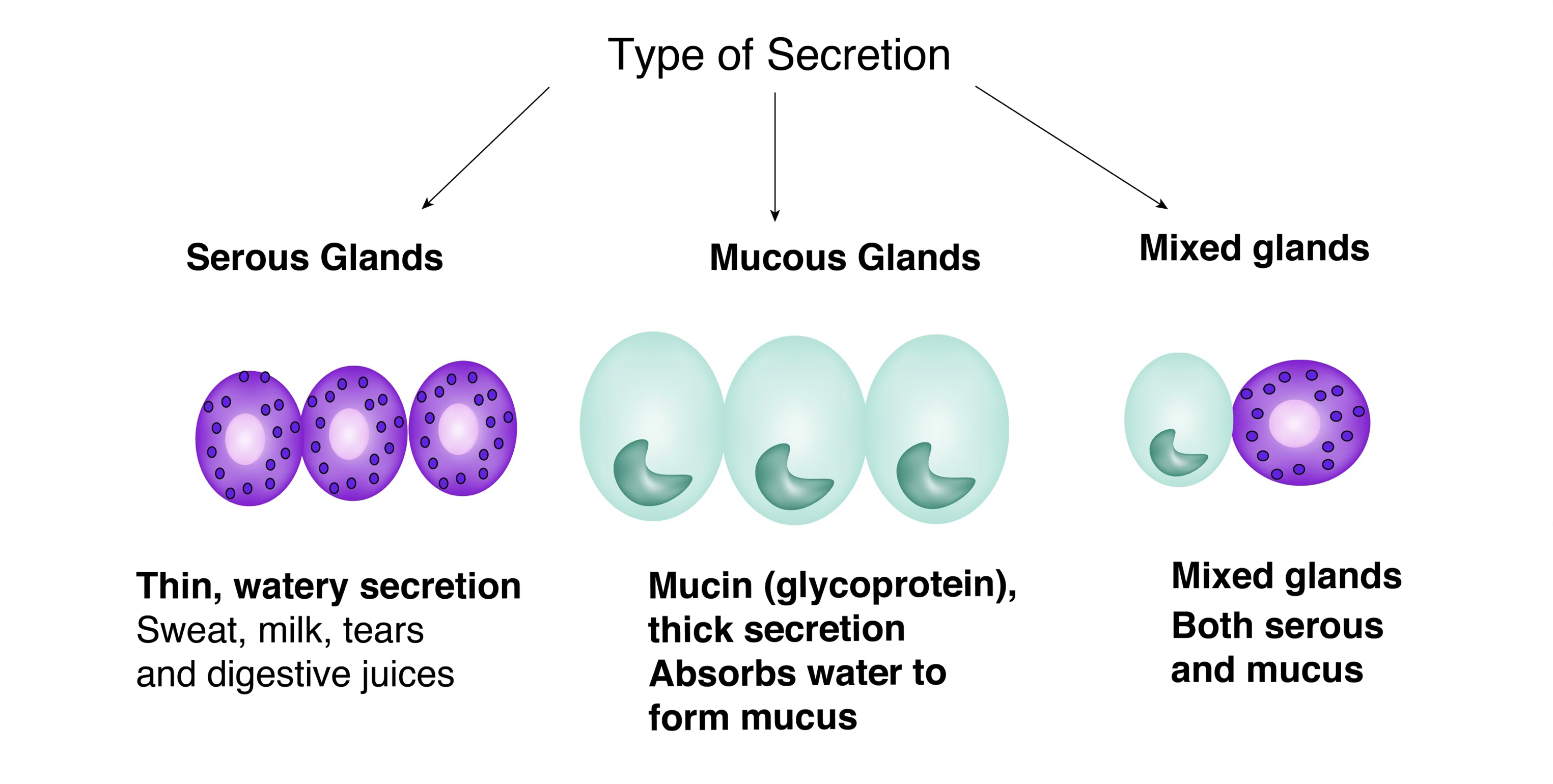
| Type | Characteristics | Example |
|---|---|---|
| Serous | Watery, protein-rich fluid | Pancreatic acini |
| Mucous | Thick, rich in glycoproteins | Goblet cells |
| Mixed | Both serous and mucous | Submandibular gland |
Serous Secrete
Serous when they secrete a watery fluid, or mucous when the secretion is thick and rich in glycoproteins.
8. Modes of Secretion
Three Mechanisms of Exocrine Secretion
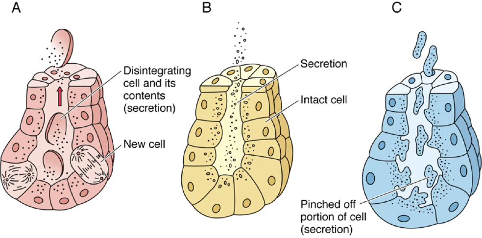
| Mode | Mechanism | Example |
|---|---|---|
| Merocrine | Pure exocytosis — no membrane or cytoplasm lost | Parotid gland |
| Apocrine | Small portion of apical cytoplasm released with product | Lactating mammary gland |
| Holocrine | Entire cell dies and becomes the secretory product | Sebaceous gland |
9. Specific Gland Types
9.1 Simple +Coiled Tubular — Sweat Glands (Merocrine)
These are simple tubular glands.
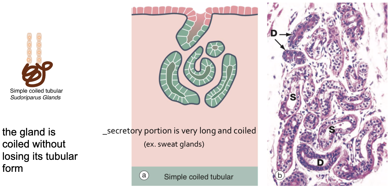
- Located deep in the dermis; composed of simple cuboidal epithelium
- Coiled duct traverses dermis and epidermis → opens at a sweat pore
- Regulated by hormones and post-ganglionic sympathetic innervation
- In women: undergo cyclical changes related to the menstrual cycle
- Secretory cells and lumina enlarge premenstrually, diminish during menstruation
Classifying 018 small intestine
https://unibo.smartzoom.com/s1241/course1776/f1791/i1792/
The epithelium is simple columnar.
There seems to be no keratin but a brushed border…
We have simple tubular glands.
Classifying 019 Large Intestine
https://unibo.smartzoom.com/s1241/course1776/f1791/i1793/
These are simple tubular cells.
qua ci ha mostrato sweat glands
e vabbe lei vede delle cose random in giro e riconosce le cose. mi sembra che i tubi fossero pluristradified cube… piu tubules of the same gland vabbe
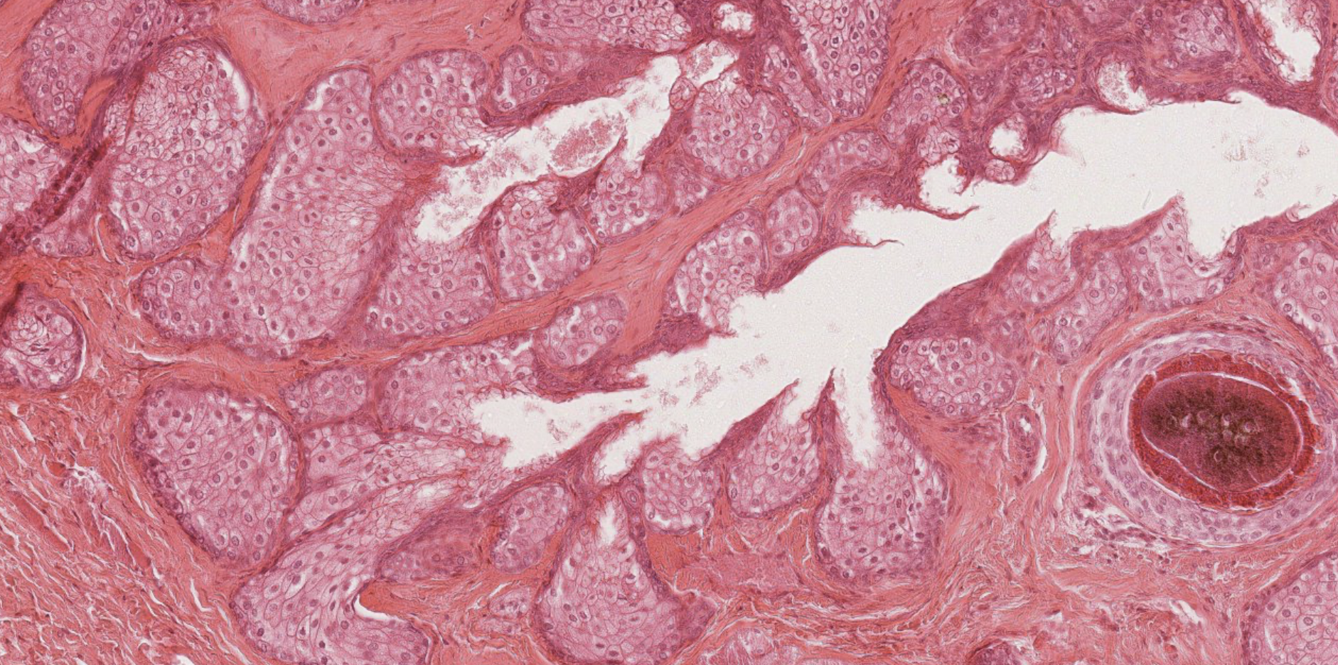
9.2 Simple Branched Acinar — Sebaceous Glands (Holocrine)
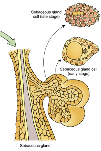
- Appendages of hair follicles; ducts open into the follicular canal
- Secretory product: sebum
Functions of Sebum
- Maintains proper skin texture and hair flexibility
- Antimicrobial
- Prevents watery fluid exchange through the skin
- Activity increases after puberty under the influence of sex hormones E CHE PALLE
non si capisce. big invagination. split in two parts. simple branched acinar gland. ok!
you do have an acinar organization of the secretory portion, but when you look atht the start of the branch, the acinar structure is lost. so this is telling you that… its a simple branched exocrine gland, you can tell that it is acinar bUT ALSO alveolar when it is functioning. okay top amo adoro. this will change the classification from acinar to alveolar… that weird duct was the… HAIR FOLLECULE WAHOOO!!!!
what are these instead? tubular coiled glands! sweat!!! wow!!!
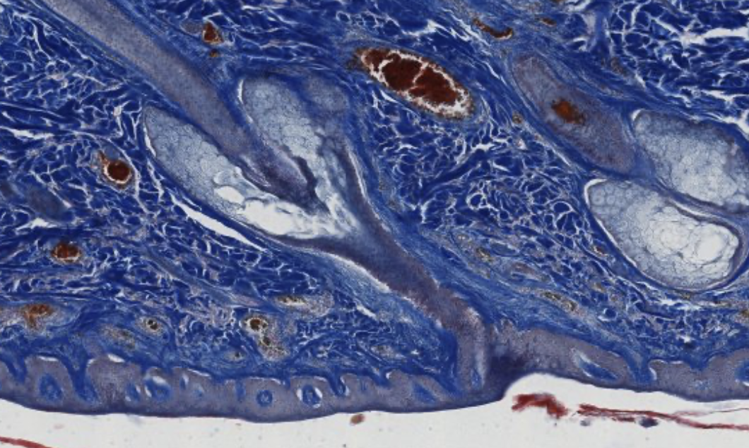
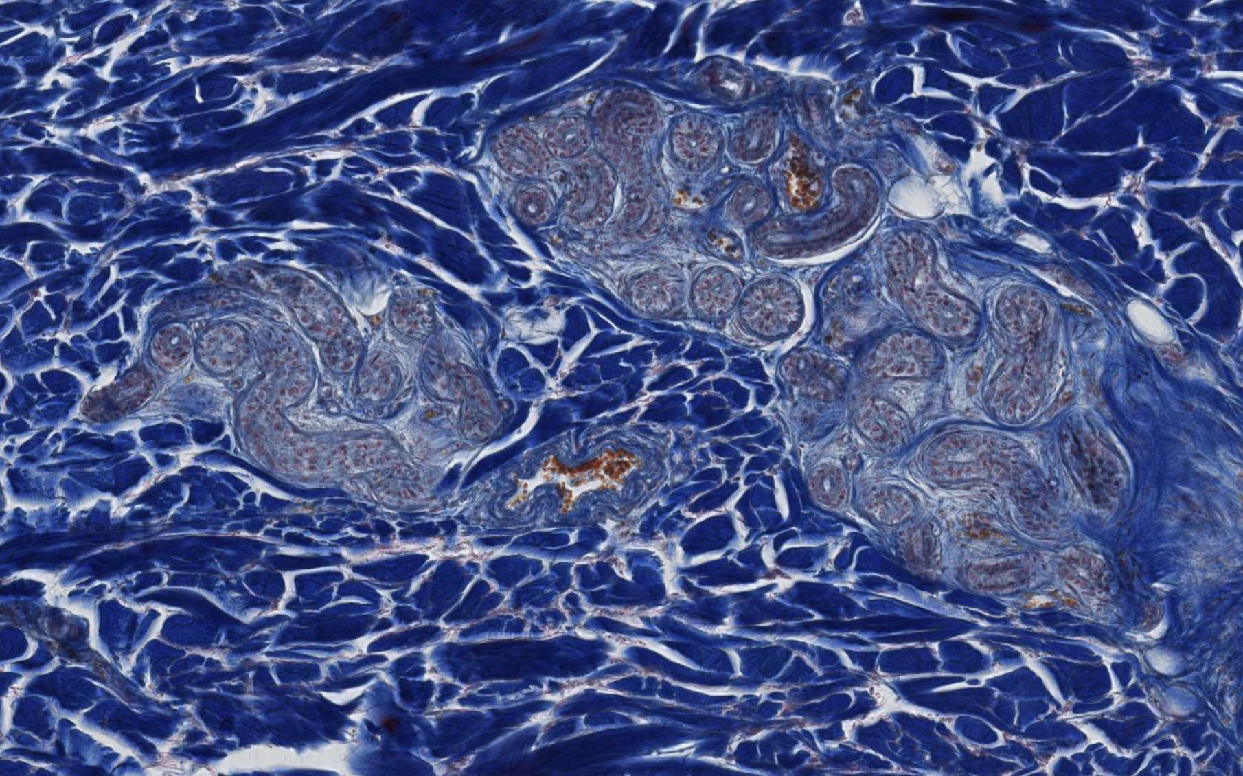
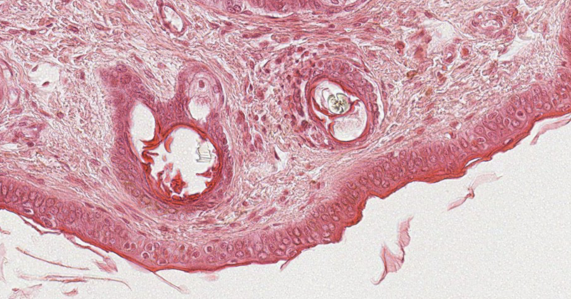
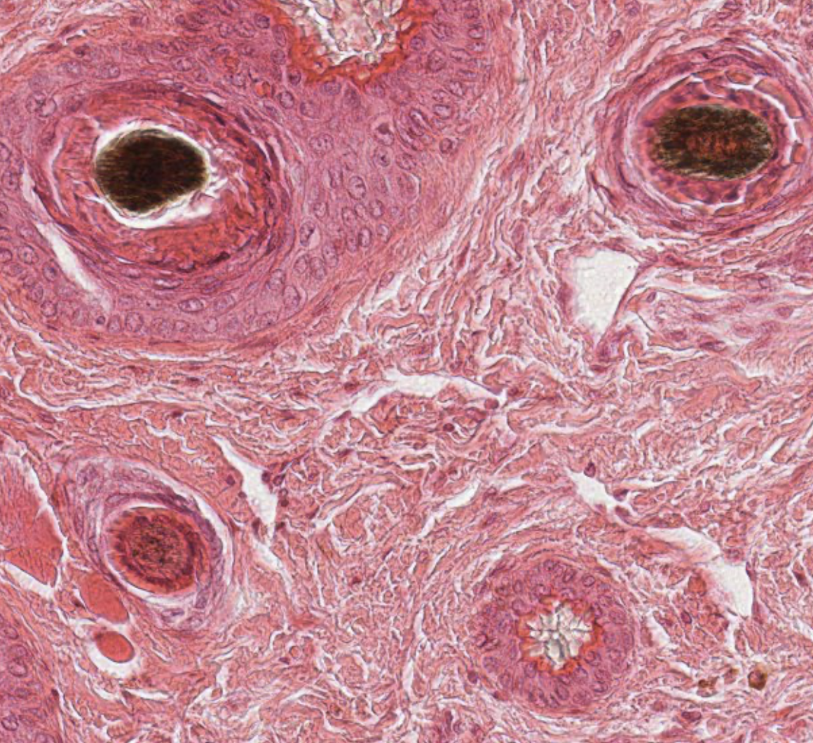
Classifying 025 eyelids
https://unibo.smartzoom.com/s1241/course1776/f1791/i1800/
stratified epithelium.
the little circles are… hair follicules! this is a cross section! you can see the epithelium surrounding the duct.
This is the important part… simple branched acinar gland (?) they’re secreting sebum, and that sebum is released through the hair follicles
Classifying...
https://unibo.smartzoom.com/s1241/course1776/f1791/i7802/
Simple branched acinar… vabbe… the secretion hasn’t started yet… non so bene il perchè…
9.3 Compound Tubulo-Acinar — Salivary Glands
Pay attention to the size of the lumen in the different ducts.
Three major salivary glands: (ordered by size >)
| Gland | Type | Notes |
|---|---|---|
| Parotid | Serous | Largest; merocrine secretion; confused often with pancreas appearance |
| Submandibular | Mixed | Serous + mucous |
| Sublingual | Mixed (65% mucous, 35% serous) | Smallest (~2–3 g); mostly mucous tubular units with serous demilunes |
 |
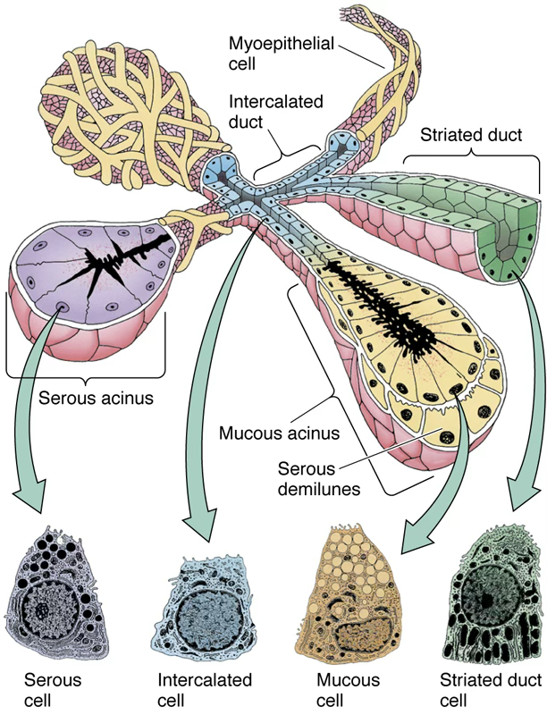
Secretory Components
- Secretory portions: serous and/or mucous acini/tubules surrounded by myoepithelial cells
- made by the compound tubul/acinar glands: the regular acinar
- in the acinar, you can’t see the lumen even if there is one (virtual lumen)
- serous secretion: incapsulated into the granule that will stay inside the cell, will get pink stained
- the nuclei occupies most of the cell body.
- for the mucus secreting unit: completely different appearence:
- single huge mucus product inside the cell: not same nucleus shape: always found in the periphery instead of the center.
- nuclei get understained because of the nature of the secrete → whiter colour
- also, myoepithelial cells cover the apical part of the mucus secreting cells.
- Duct system: (>1) intercalated ducts
- interlobular duct: gfarest area of the secretory portion, two of the epithelia: columnar, stratified columnar / pseudo stratified, collector duct with huge lumen
- striated duct: simple epithelium, one layer of cuboidar / columnar. striation inside the cells. intercalated in the
- intercaleted duct: collects the secrete directly from the secretory portion.
- Protective secretions: lysozyme, lactoferrin, secretory IgA
- Huge Parenchima
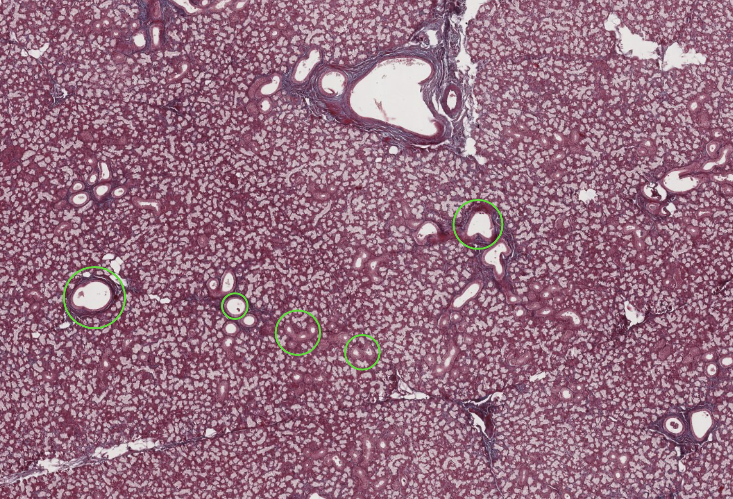
Classifying 030 salivary gland
Most of the sample is occupied by glands. Huge network of ducts. Obv compound exocrine gland…
The compound marker is the number of ducts. Cuboidal epithelium… idk where
Finding holes → ducts → classifying by the diameter… we mainly see terminal ducts, the biggest.
The nuclei helps us classify what secreting portion this is.
When its white and elongated, nucleus at the periphery… mucus portion. When its small circles full of cells with nucleus at the center and dark staining… acinar…
Be careful of the cut when seeing ducts.
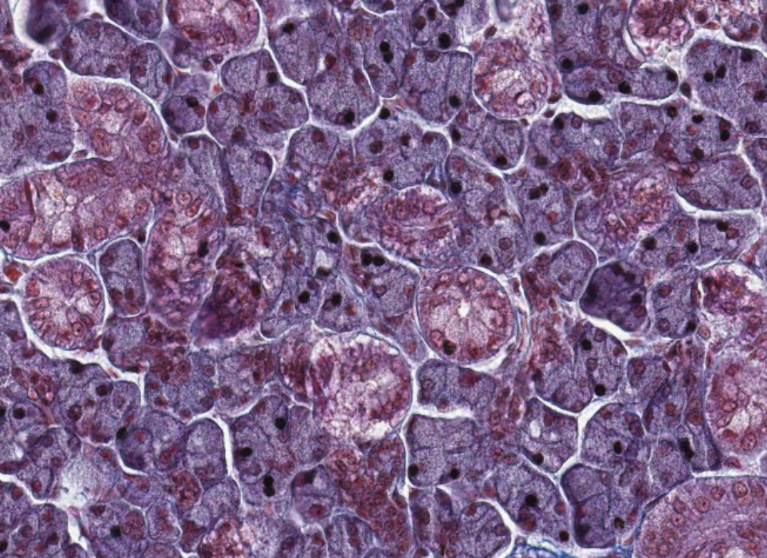
Classifying 028 parotid
Several ducts, surely compound exocrine."Very Pretty" - Prof. Lauriola
Secretory portion…
Has to be serous acinar…
Here we have only that type!
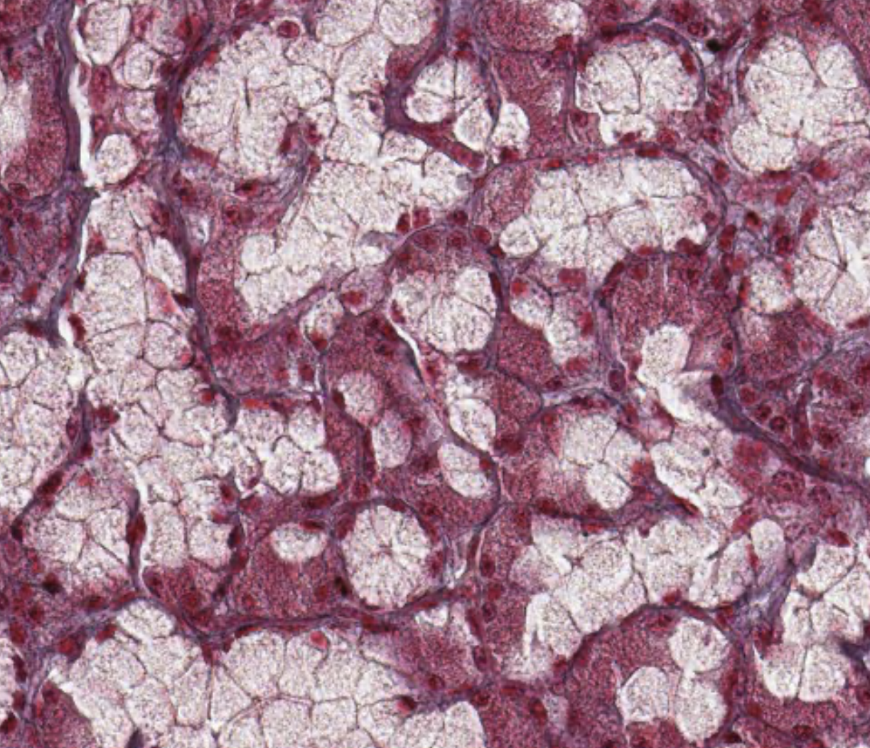
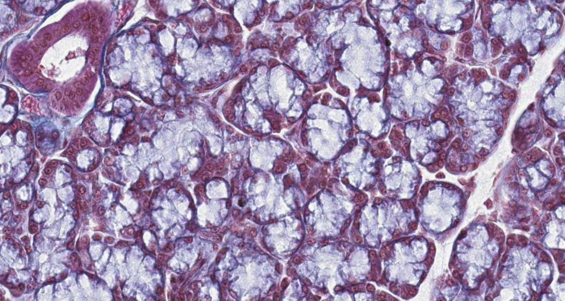
Classifying 031
Due to the number of ducts, surely exocrine compound.
Time to check for the secretory portions:
We can see some acinar and some nucleus in periphery ligher ones so also tubular mucus secretion.
Here its almost all mucus secreting.
9.4 Compound Acinar — Exocrine Pancreas (Serous)
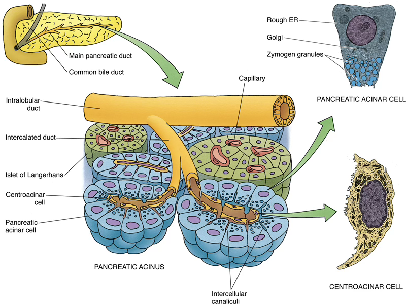
Acinar Cell Products
Enzyme Function Pancreatic amylase Breaks down starches, carbohydrates, glycogen Pancreatic lipase Breaks down fats Cholesterol esterase Breaks down cholesterol esters → fatty acids DNase / RNase Break down DNA / RNA Elastase Breaks down elastic fibers Trypsin / Chymotrypsin Break down proteins → short peptides Trypsin inhibitor Protects cells from intracellular trypsin activation
- Lumen of each acinus is occupied by centroacinar cells (beginning of the duct system)
- Centroacinar cells produce a bicarbonate-rich buffer:
- Release stimulated by secretin (from DNES Diffuse NeuroEndocrine Cells of small intestine) and acetylcholine
- Duct hierarchy: centroacinar (low cuboidal centroacinar cells) → intercalated → intralobular → interlobular ducts
Dual Regulation
Enzyme-rich (acinar) and enzyme-poor (centroacinar/bicarbonate) secretions are regulated separately and may be released at different times or simultaneously.
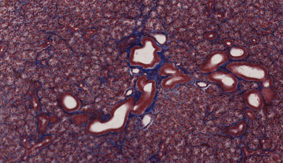
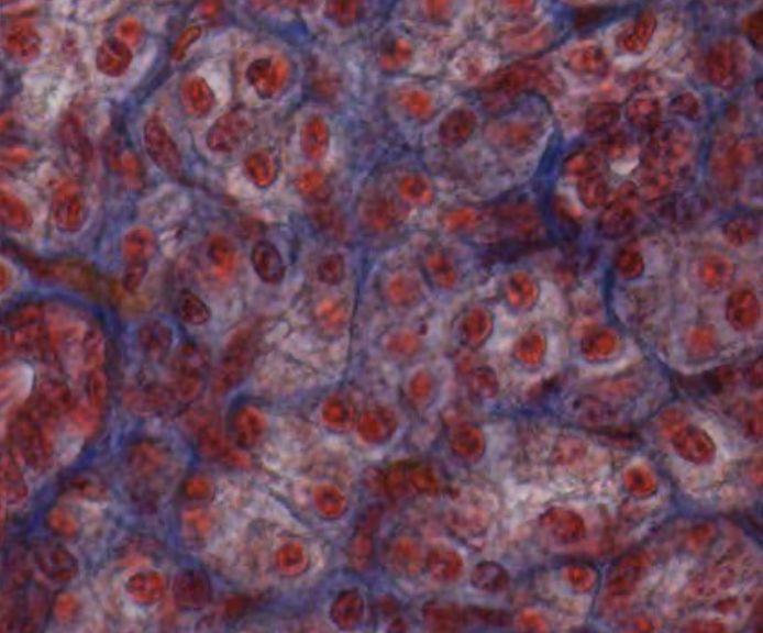
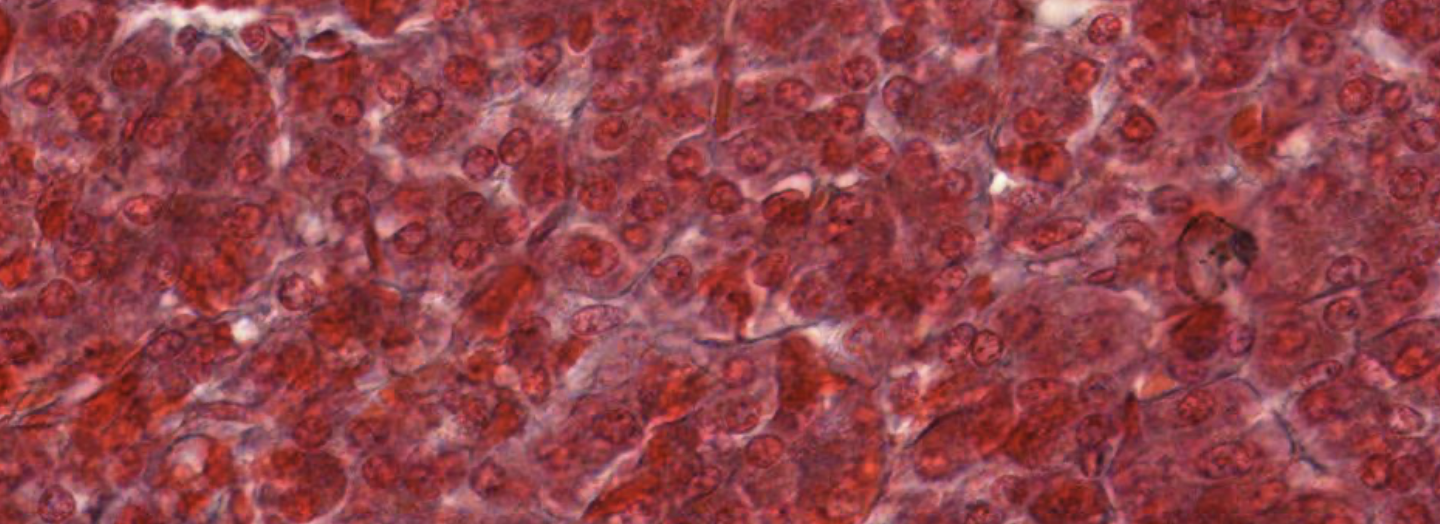
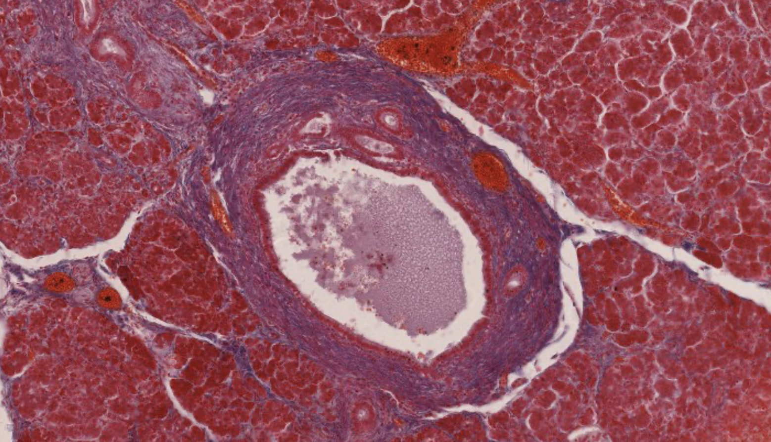
Classifying 029 pancreas
Interlobular duct… connecting more than one duct. Epithelium is simple columnar, the pancreas is surrounded by a capsule that divides it into lobi.
Acinar units, the nuclei are rounded, with a huge amount of enzymes at the center.
There are some arteries, that cant be classified as ducts since no columnar epithelium + theres muscle cells present.
Acinar cell surrounded by reticular something…
It’s hard to catch the duct due to how the cut is made.
The endocrine component is the marker of the pancreas → there are langerhans cells.
Interlobular duct, the biggest.

9.5 Compound Tubulo-Alveolar — Mammary Gland (Apocrine + Merocrine)
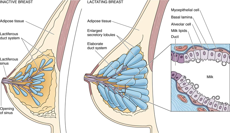
- Composed of 15–20 lobes radiating from the nipple, each drained by a lactiferous duct
- Each lactiferous duct dilates to form a lactiferous sinus (milk storage) before narrowing at the nipple
- Mammary glands can be found as resting/nonsecreting when the woman is not in pregnancy and lactating/active.
Milk composition:
- Proteins, lipids, lactose
- Lymphocytes, monocytes, antibodies
- Minerals and fat-soluble vitamins
Hormonal Regulation
Hormone Role Estrogen + Progesterone Activate gland during pregnancy; stimulate alveolar development Prolactin (from anterior pituitary acidophils) Activates milk secretion after birth, when E/P levels drop Colostrum Protein-rich fluid produced before milk; present at end of pregnancy
Dual secretory mechanism of alveolar cells:
- Lipids → released by apocrine exocytosis
- Proteins → released by merocrine exocytosis
Classifying 032 mammary gland
This is one of the main ducts,
apocrine… alveolar… non capisco. some are “closed” still havent secreted because the shape and size of cell → much taller.
nonn ho capito niente di questo
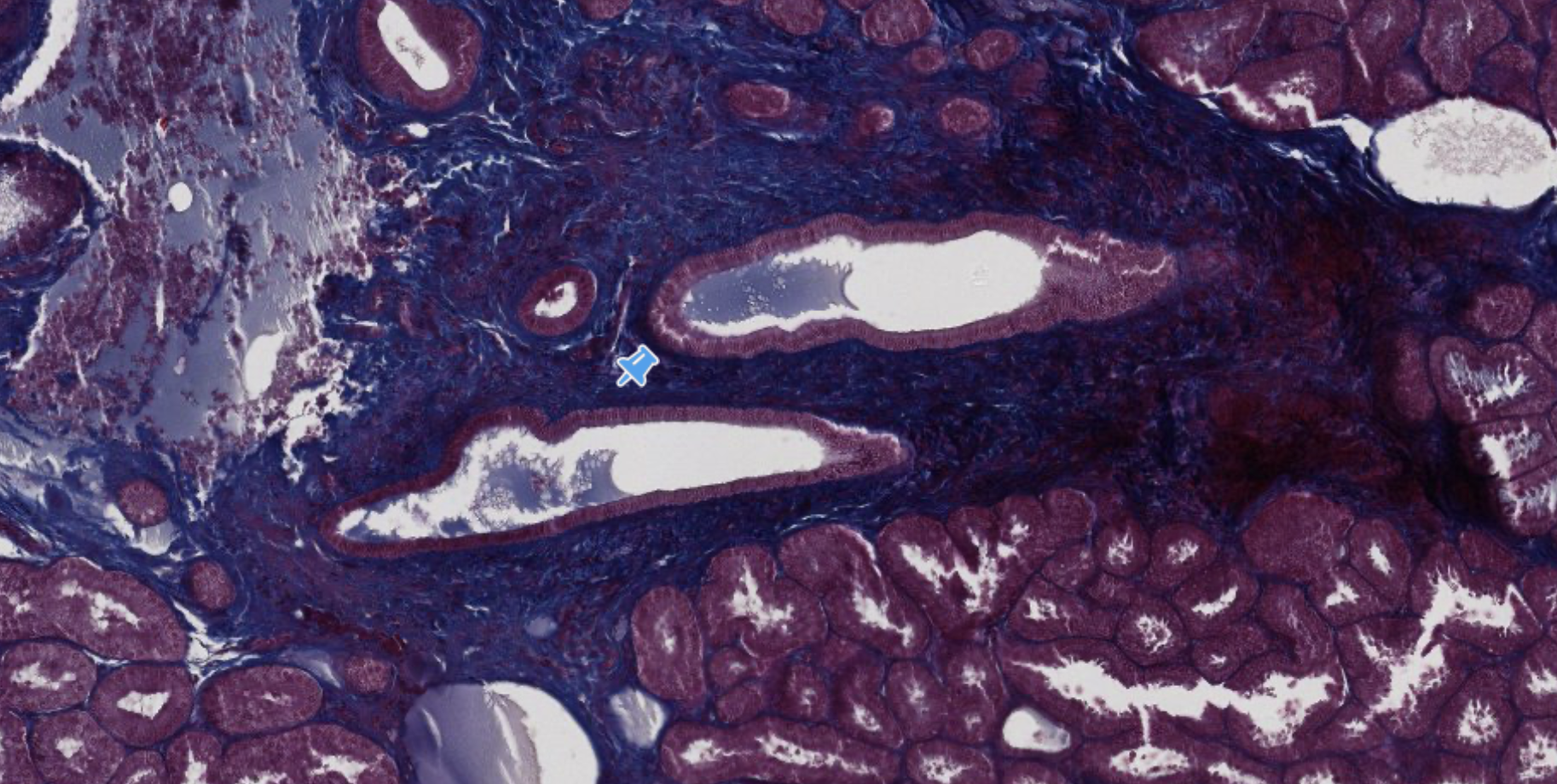
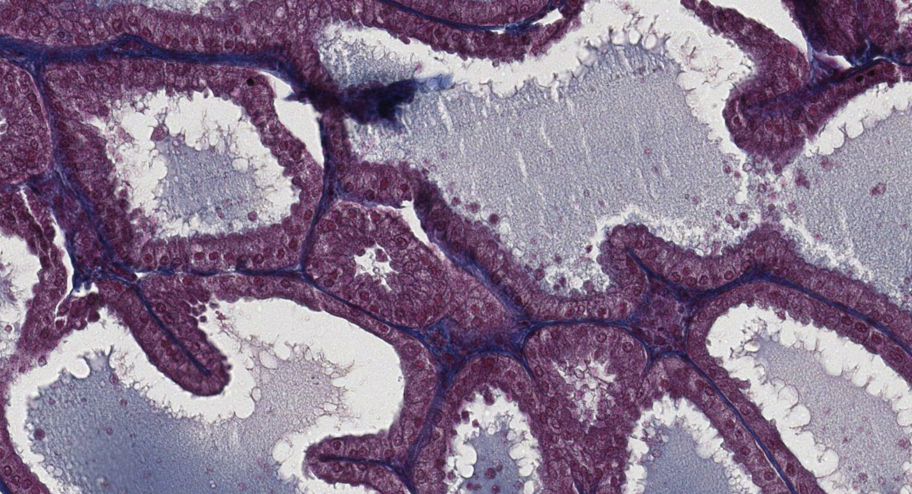
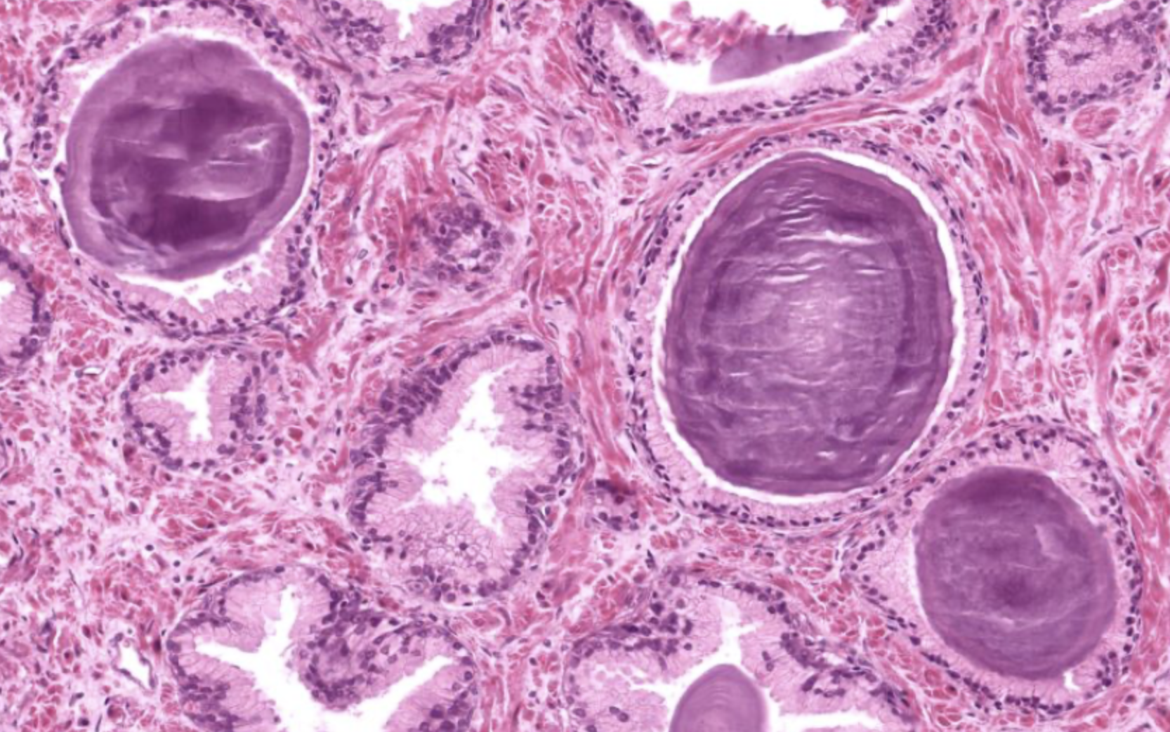
9.6 Compound Tubulo-Alveolar — Prostate Gland
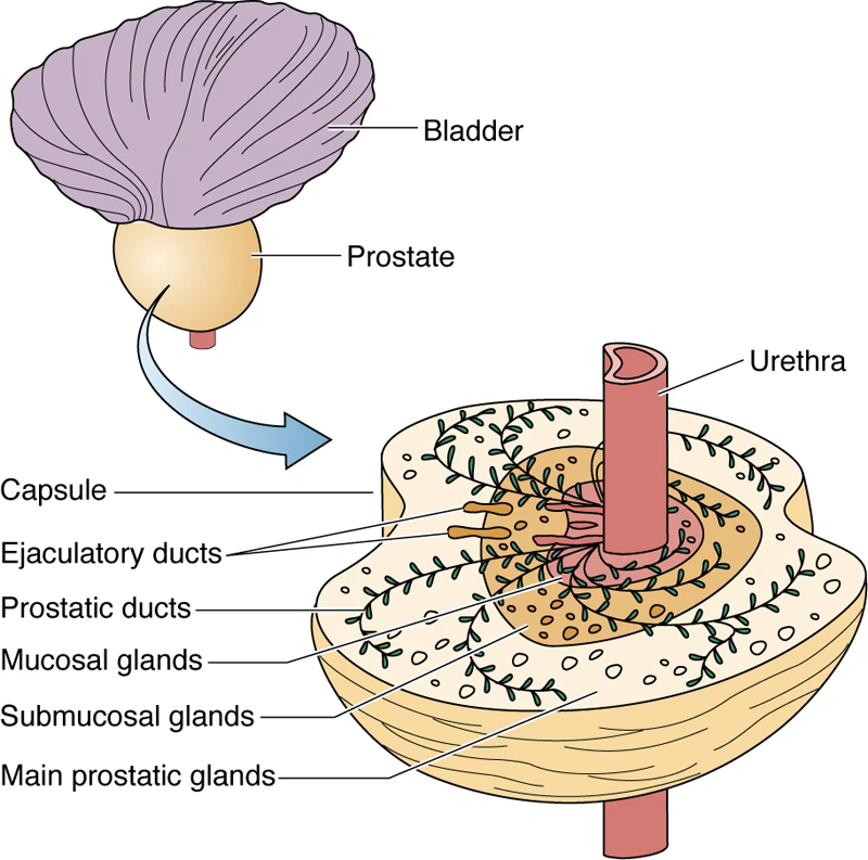
- Largest male accessory gland; pierced by the urethra and ejaculatory ducts
- Capsule: dense irregular collagenous connective tissue with smooth muscle cells
- Consists of 30–50 individual compound tubuloalveolar glands in 3 concentric layers:
- Mucosal
- Submucosal
- Main
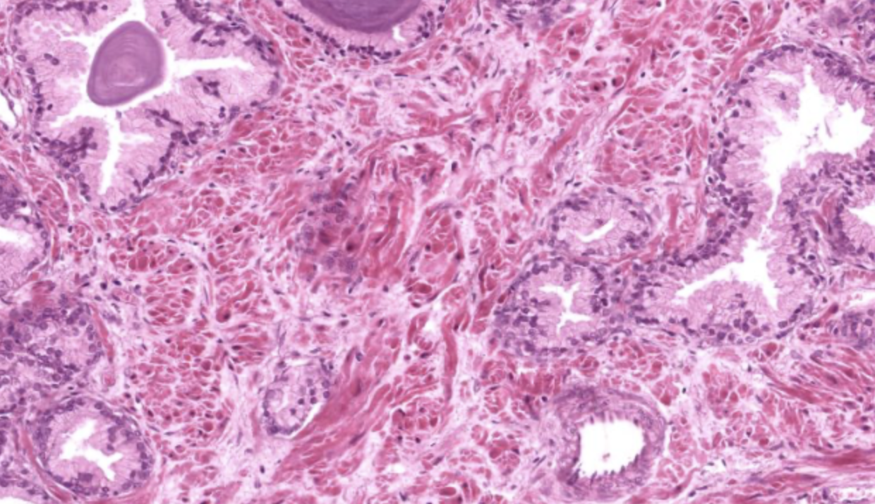
Prostatic Concretions
Lumina frequently display corpora amylacea — calcified glycoprotein concretions that increase in number with age.
Prostatic secretion (part of semen):
- Serous, white fluid
- Rich in: lipids, proteolytic enzymes, acid phosphatase, fibrinolysin, citric acid
- Regulated by dihydrotestosterone (DHT) — the active form of testosterone
Example
Alveolar organization. Type of secretion? only merocrine, so we’re not gonna lose the apical domain
The big dark circles are the corpora amylacea, main indicator that we’re looking at a prostate:
9.7 Liver (Endocrine + Exocrine)
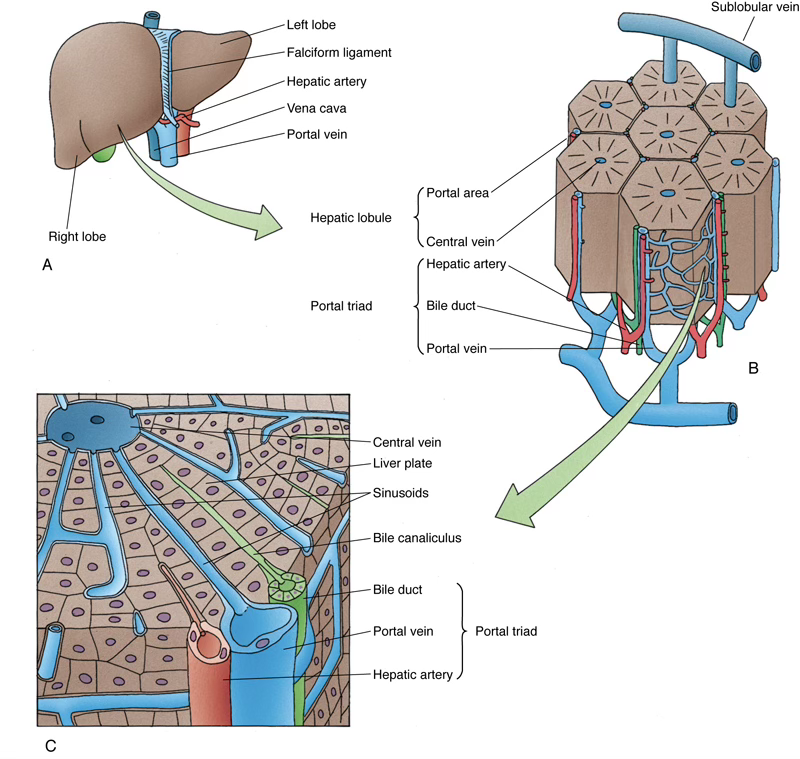
Dual Function
The same cell — the hepatocyte — is responsible for both:
- Exocrine function: bile production
- Endocrine function: plasma protein synthesis, glucose regulation, etc.
- Also: detoxification of toxins → excreted in bile
Structural organization:
| Structure | Description |
|---|---|
| Glisson’s capsule | Connective tissue capsule; enters at porta hepatis |
| Portal triad | 1 portal vein + 1 hepatic artery + 1 bile duct |
| Classical lobule | Hexagonal unit; hepatocytes arranged in plates |
| Central vein | Receives blood from all sinusoids of the lobule |
Blood supply:
- 25% — oxygenated blood via hepatic arteries
- 75% — nutrient-rich blood via portal vein
Hepatocyte plasma membrane domains:
| Domain | Feature |
|---|---|
| Lateral | Forms bile canaliculi (1–2 μm diameter) → conducts bile to lobule periphery |
| Sinusoidal | Has microvilli projecting into the space of Disse |
| ![[Pasted image 20260319104726.png | 400]] |
| Hepatocyte organelles: |
- Abundant free ribosomes, RER, Golgi apparatus
- Up to 2000 mitochondria per cell
- Inclusions: lipid droplets and glycogen
Classifying This
Bel sito ha tutti i bottoni per andare alle diverse strutture wooo!!!
TLDR
TLDR — Epithelium and Glands
Glands are secretory structures classified as endocrine (ductless, secrete into blood/lymph) or exocrine (use ducts to secrete onto surfaces).
Exocrine glands are further classified by:
- Morphology: simple vs. compound; tubular vs. acinar vs. tubulo-acinar
- Secretion type: serous (watery), mucous (thick, glycoprotein-rich), or mixed
- Mode of secretion:
- 🔵 Merocrine = pure exocytosis (e.g., parotid, sweat glands)
- 🟡 Apocrine = apical cytoplasm lost with product (e.g., mammary lipid secretion)
- 🔴 Holocrine = entire cell becomes product (e.g., sebaceous glands)
Key glands to know:
| Gland | Type | Mode | Key Features |
|---|---|---|---|
| Sweat gland | Simple coiled tubular | Merocrine | Sympathetic control; menstrual cycle variation |
| Sebaceous gland | Simple branched acinar | Holocrine | Sebum; sex hormone-dependent |
| Salivary glands | Compound tubulo-acinar | Merocrine | Serous (parotid), mixed (submandibular/sublingual) |
| Exocrine pancreas | Compound acinar | Merocrine | Digestive enzymes + bicarbonate buffer; secretin regulated |
| Mammary gland | Compound tubulo-alveolar | Apocrine (lipids) + Merocrine (proteins) | Estrogen/progesterone → development; prolactin → milk |
| Prostate | Compound tubulo-alveolar | Merocrine | DHT-regulated; corpora amylacea increase with age |
| Liver | Compound (hepatocyte plates) | Merocrine | Dual endocrine/exocrine; portal triad; bile canaliculi; space of Disse |
Signaling distances: autocrine (self) → paracrine (nearby) → endocrine (distant via blood).
Unicellular glands: goblet cells secrete mucinogen via exocytosis triggered by chemical irritation or parasympathetic stimulation.