TARGET DECK: MED::I::Morphology and Development::Histology::11 - Nervous Tissue
Overview
Key Facts
- One of the four main tissues of the body
- Ectodermal origin (neural crest cells)
- Main function: communication
Composition
| Component | Role | Size Range |
|---|---|---|
| Neurons | Reception & transmission of impulses | – |
| Neuroglia | Physical & metabolic support | — |
Anki cloze
Nervous tissue has {1:ectodermal (neural crest cell)} origin and its main function is {1:communication}.
Anki cloze
Neurons range in size from {1:5 µm} (smallest) to {1:150 µm} (largest).
Anatomical Organization
| Division | Structures |
|---|---|
| CNS | Brain + spinal cord |
| PNS | Cranial & spinal nerves + ganglia |
- PNS has afferent (sensory) and efferent (motor) components
Anki cloze
The CNS consists of the {1:brain and spinal cord}; the PNS consists of {1:cranial and spinal nerves and ganglia}.
Neurons
Definition
Neurons are the cells responsible for the reception and transmission of nerve impulses to and from the CNS.
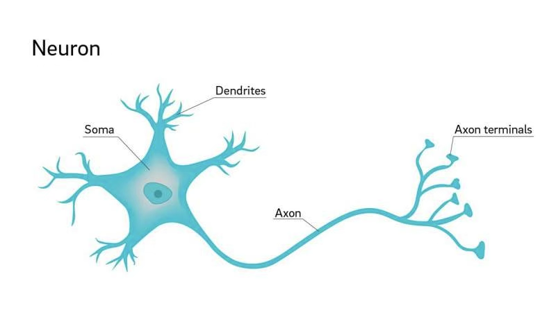
Parts of a Neuron
Most neurons are composed of three distinct parts:
- Cell body (soma / perikaryon)
- Multiple dendrites
- A single axon
Anki cloze
The three main parts of a neuron are: {1:cell body (soma)}, {1:dendrites}, and {1:a single axon}.
Cell Body (Soma / Perikaryon)
- Perikaryon = Greek peri (around) + karyon (nucleus)
- Contains the nucleus and surrounding cytoplasm
- Specialized for reception and integration of information
- The cell body is the most conspicuous region, but the largest volume of cytoplasm is actually in the processes
Nucleus
- Large, usually spherical, centrally located
- Finely dispersed chromatin → rich synthetic activity
- Smaller neurons may have condensed, inactive heterochromatin
- Well-defined nucleolus commonly present
Cytoplasm (Ultrastructure)
- Abundant rough endoplasmic reticulum (RER) with parallel cisternae arrays — especially prominent in large motor neurons
- Polyribosomes scattered throughout cytoplasm
- Stacked RER cisternae + polyribosomes stain with basic dyes → basophilic clumps = Nissl bodies (visible under LM)
- RER present in dendritic regions (scattered short/branching cisternae)
- RER absent at the axon hillock — only SER is present in the axon
Nissl Bodies
Nissl bodies are clumps of RER + polyribosomes in the soma and dendrites. They are absent at the axon hillock — this is a key LM landmark.
Anki cloze
Nissl bodies are formed by stacked {1:rough endoplasmic reticulum cisternae} and {1:polyribosomes} stained with basic dyes.
Anki cloze
RER (Nissl substance) is absent at the {1:axon hillock}; only {1:smooth ER} is present in the axon.
Dendrites
- Projections from the cell body specialized for receiving stimuli from sensory cells, axons, and other neurons
- Often multibranched (arborized) → receive multiple stimuli simultaneously
- Impulses received by dendrites are transmitted toward the soma
- Form a dendritic tree collectively
- Covered by small protrusions: dendritic spines → establish axonal synaptic connections
Anki cloze
Dendritic spines establish {1:axonal synaptic connections}.
Axon
- Each neuron has a single axon
- Varying diameter; up to 100 cm in length
- Conducts impulses away from the soma to other neurons, muscles, or glands
- May also receive stimuli from other neurons, modifying its behavior
- Originates from the soma at the axon hillock
- Ends in a terminal arborization = telodendron
- Each terminal branch of the telodendron has an enlarged ending: synaptic terminal or synaptic bouton
- Axon terminals (end bulbs) approach other cells to form synapses
Anki cloze
The axon originates from the soma at the {1:axon hillock} and terminates in the {1:telodendron}, whose enlarged endings are called {1:synaptic boutons}.
Anki cloze
The axon conducts impulses {1:away from} the soma; dendrites conduct impulses {1:toward} the soma.
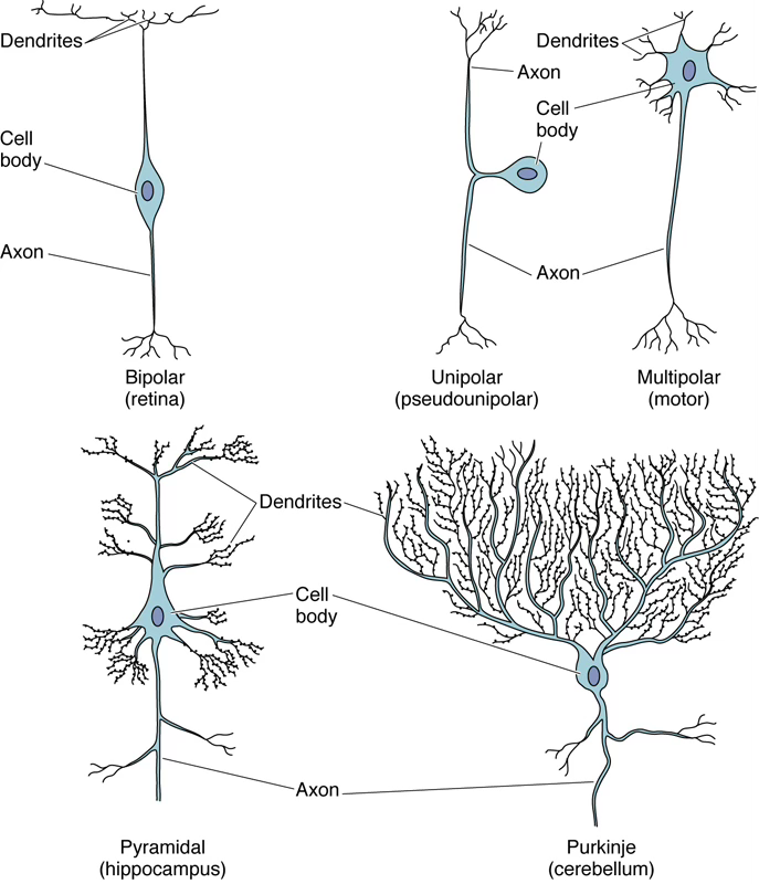
Neuron Classification
Morphological Classification
| Type | Processes | Location / Example |
|---|---|---|
| Multipolar | 1 axon + ≥2 dendrites | Motor neurons, pyramidal cells (cerebral cortex), Purkinje cells (cerebellum) — most common |
| Bipolar | 1 axon + 1 dendrite | Olfactory epithelium, retina, inner ear |
| Pseudounipolar | 1 axon splitting into 2 branches | Sensory ganglia of PNS (dorsal root ganglion) |
Pseudounipolar Detail
The single axon splits into:
- One central process (→ CNS)
- One peripheral process (from sensory endings)
Impulses generated in sensory endings travel through both branches to the CNS.
Anki cloze
The most common type of neuron is the {1:multipolar} neuron, which has one axon and {1:two or more} dendrites.
Anki cloze
Bipolar neurons are found in specialized tissues such as the {1:olfactory epithelium}, {1:retina}, and {1:inner ear}.
Anki cloze
Pseudounipolar neurons are found in the {1:sensory ganglia of the PNS (dorsal root ganglia)}.
Functional Classification
| Type | Direction | Examples |
|---|---|---|
| Motor neurons (efferent) | CNS → effectors | Somatic efferent, visceral efferent |
| Sensory neurons (afferent) | Receptors → CNS | Somatic afferent, visceral afferent |
| Interneurons (intercalated) | Between sensory & motor | Communicating/integrating network |
Anki cloze
Interneurons form a {1:communicating and integrating} network between {1:sensory and motor} neurons.
Neuroglia (“Nerve Glue”)
Info
Neuroglia are 10× more common than neurons. Main function: support.
Summary Table
| Cell | Location | Function |
|---|---|---|
| Satellite cells | PNS (ganglia) | Surround neuron cell bodies; maintain controlled microenvironment; regulate , , nutrients, neurotransmitter levels |
| Schwann cells | PNS (nerve fibers) | Myelinate PNS axons (1 Schwann cell : 1 axon); guide axon regrowth; form neurolemma |
| Oligodendrocytes | CNS | Myelinate CNS axons (1 oligodendrocyte : multiple axons); structural support |
| Astrocytes | CNS | Support/brace neurons; anchor to blood supply; guide migration; potassium spatial buffering; regulate ion/nutrient/gas concentrations; absorb & recycle neurotransmitters; form scar tissue |
| Microglia | CNS | Phagocytosis of debris, pathogens, dead cells; maintained by self-renewal |
| Ependymal cells | CNS (ventricles & central canal) | Line ventricles (brain) and central canal (spinal cord); assist CSF production & circulation; have motile cilia |
Key Comparison: Myelination
- Schwann cells (PNS): 1 cell myelinates 1 internode of 1 axon
- Oligodendrocytes (CNS): 1 cell myelinates internodes of multiple axons
Anki cloze
In the PNS, myelination is performed by {1:Schwann cells}; in the CNS, by {1:oligodendrocytes}.
Anki cloze
A single oligodendrocyte can myelinate {1:multiple axons}, whereas a single Schwann cell myelinates only {1:one axon}.
Anki cloze
Astrocytes regulate extracellular potassium concentration through a process called {1:potassium spatial buffering}.
Anki cloze
Microglia maintain their population by {1:self-renewal} and are functionally/developmentally {1:unrelated to monocytes}.
Anki cloze
Ependymal cells line the {1:ventricles of the brain} and the {1:central canal of the spinal cord} and bear {1:motile cilia}.
Astrocyte Subtypes
| Subtype | Location |
|---|---|
| Fibrous astrocytes | White matter |
| Protoplasmic astrocytes | Gray matter |
Anki cloze
Fibrous astrocytes are found in {1:white matter}; protoplasmic astrocytes are found in {1:gray matter}.
Shared Functions (CNS ↔ PNS Analogues)
| Function | PNS Cell | CNS Cell |
|---|---|---|
| Myelination | Schwann cells | Oligodendrocytes |
| Ion/nutrient/gas regulation | Satellite cells | Astrocytes |
Myelin Sheath
Structure
- Myelin = plasmalemma of the Schwann cell (or oligodendrocyte) wrapped multiple times around the axon
- Prevents leakage of the action potential
- EM: alternating lines at intervals:
- Major dense line — fused cytoplasmic surfaces of the Schwann cell membrane
- Intraperiod line — apposing outer leaflets of the Schwann cell membrane
Anki cloze
The major dense line of myelin represents the fused {1:cytoplasmic surfaces} of the Schwann cell plasma membrane.
Myelin Formation
- Oligodendrocyte (or Schwann cell) concentrically wraps its membrane around the axon
- Wrapping may continue for >50 turns
- During wrapping, cytoplasm is squeezed back → cytoplasmic surfaces contact each other → major dense line
Developmental Note
- Motor nerves: nearly completely myelinated at birth
- Sensory roots: myelinated several months after birth
- Some CNS tracts: not fully myelinated until several years after birth
- Nerves are not myelinated simultaneously during development
Unmyelinated Nerve Fibers
- Predominate in gray matter; axons are thin
- In PNS: a single Schwann cell houses several unmyelinated axons in individual cytoplasmic invaginations — no myelin produced
- Entire axolemma is freely exposed to interstitial tissue; partially protected by a basal lamina surrounding the Schwann cell
Conduction Speed Comparison
| Type | Mechanism | Max Speed |
|---|---|---|
| Myelinated | Saltatory conduction (node to node) | |
| Unmyelinated | Continuous conduction |
Anki cloze
Myelinated nerves conduct impulses via {1:saltatory conduction} at up to {1:120 m/s}; unmyelinated nerves conduct continuously at up to {1:15 m/s}.
Peripheral Nerves
Connective Tissue Investments
Peripheral nerves are bundles of nerve fibers (axons) surrounded by three connective tissue sheaths:
| Layer | Coverage | Composition |
|---|---|---|
| Epineurium | Entire nerve | Dense irregular collagenous CT; type I collagen + fibroblasts; contains arteries, veins, lymphatics |
| Perineurium | Each fascicle | Dense CT (thinner than epineurium); several concentric layers of neuroepithelial perineurial cells joined by tight junctions → forms blood-nerve barrier; basal lamina of type IV collagen + laminin |
| Endoneurium | Individual axons | Type III collagen fibrils; few fibroblasts, macrophages, mast cells, endoneurial capillaries |
Blood-Nerve Barrier
The perineurium is responsible for maintaining the homeostatic microenvironment of the endoneurium via tight junctions between perineurial cells.
Mnemonic — Nerve Layers (outside → in)
“Every Penguin Enjoys” = Epineurium → Perineurium → Endoneurium
Anki cloze
From outermost to innermost, the connective tissue layers of a peripheral nerve are: {1:epineurium}, {1:perineurium}, and {1:endoneurium}.
Anki cloze
The blood-nerve barrier is formed by {1:tight junctions} between {1:perineurial cells} of the perineurium.
Anki cloze
The endoneurium contains {1:type III collagen} and surrounds {1:individual axons and their Schwann cells}.
- Virtual microscopy links (UniBo):
- https://virtualmicroscopy.patologia-sperimentale.unibo.it/_contenuti/index.php?viewPage=9&bttl=2&lingua=ITA&page=2&cell_id=333
- https://virtualmicroscopy.patologia-sperimentale.unibo.it/_contenuti/index.php?viewPage=9&bttl=2&lingua=ITA&page=2&cell_id=349
- https://www.histologyguide.com/quizzes/06-nervous-tissue.html#top-04
Synapses
Definition
Synapses are sites where nerve impulses are transmitted from a presynaptic cell to a postsynaptic cell. Transmission can be electrical or chemical; chemical synapses are the most common.
Types of Synaptic Contacts
| Type | Connection |
|---|---|
| Axodendritic | Axon → dendrite |
| Axosomatic | Axon → soma |
| Axoaxonic | Axon → axon |
| Dendrodendritic | Dendrite → dendrite |
Anki cloze
The four main types of synaptic contacts are: {1:axodendritic}, {1:axosomatic}, {1:axoaxonic}, and {1:dendrodendritic}.
Structure of the Synapse
- Presynaptic terminal (bouton terminal): bulbous expansion at the axon end
- Presynaptic cytoplasm contains:
- Mitochondria
- Elements of smooth ER
- Abundance of synaptic vesicles (– diameter) filled with neurotransmitter
- Peptide neurotransmitters are manufactured in the cell body and transported to the terminal via anterograde transport
- Non-peptide neurotransmitters are manufactured and packaged near the axon terminal
- Enzymes in axoplasm protect neurotransmitters from degradation
- Cone-shaped presynaptic densities project from the membrane into the cytoplasm → form the active site
- A reserve pool of synaptic vesicles adheres to actin microfilaments
- Cell adhesion molecules (CAMs) act as signaling molecules at both pre- and postsynaptic membranes
Anki cloze
Synaptic vesicles are {1:40–60 nm} in diameter and are filled with {1:neurotransmitter}.
Anki cloze
Peptide neurotransmitters are synthesized in the {1:cell body} and transported to the axon terminal by {1:anterograde transport}.
Mechanism of Neurotransmitter Release
- Action potential reaches the presynaptic membrane
- Voltage-gated channels open → enters
- influx causes synaptic vesicles (under SNARE protein influence) to fuse with the presynaptic membrane
- Neurotransmitter released into the synaptic cleft via exocytosis
- Excess membrane recaptured via clathrin-mediated endocytosis
- Endocytic vesicle fuses with smooth ER → membrane recycled
Anki cloze
Synaptic vesicle fusion with the presynaptic membrane is triggered by {1:Ca²⁺ influx} and mediated by {1:SNARE proteins}.
Anki cloze
After exocytosis, excess presynaptic membrane is recaptured by {1:clathrin-mediated endocytosis}.
Postsynaptic Events
- Postsynaptic membrane contains neurotransmitter receptors (ligand-gated ion channels)
- Neurotransmitter binding → ion channel opening → altered membrane permeability → reversal of membrane potential
- Depolarization = excitatory response
- Hyperpolarization = inhibitory response
- Glial cells increase synaptogenesis, synaptic efficacy, and action-potential firing
Anki cloze
Binding of a neurotransmitter to postsynaptic receptors causes either {1:depolarization} (excitatory) or {1:hyperpolarization} (inhibitory) of the postsynaptic membrane.
Autonomic Nervous System (ANS)
Overview
The ANS is a motor system controlling viscera by innervating smooth muscle, cardiac muscle, and glands.
Unlike the somatic system (1 neuron: CNS → effector), the ANS has 2 neurons between CNS and effector organ.
Divisions of the ANS
| Feature | Sympathetic | Parasympathetic |
|---|---|---|
| Physiological role | ”Fight or flight” | Homeostasis |
| Heart rate | ↑ | ↓ |
| Blood pressure | ↑ | ↓ |
| Respiration | ↑ | ↓ |
| Blood flow to skeletal muscle | ↑ | ↓ |
| Pupils | Dilated | Constricted |
| Visceral function | ↓ | ↑ |
| Postganglionic neurotransmitter | Noradrenaline (adrenergic) | Acetylcholine (cholinergic) |
Anki cloze
The sympathetic nervous system prepares the body for {1:“fight or flight”}; the parasympathetic nervous system promotes {1:homeostasis}.
Anki cloze
In contrast to the somatic motor system (1 neuron CNS → effector), the ANS uses {1:two neurons} between the CNS and the effector organ.
CNS Organization
Gray Matter vs. White Matter
| Gray Matter | White Matter | |
|---|---|---|
| Contents | Neuronal cell bodies | Myelinated axon fibers |
| Location in cerebrum | Cerebral cortex (outermost layer) | Inside |
| Location in spinal cord | Inside (H-shape) | Outside |
Brain Regions
- Cerebral cortex: contains several layers of pyramidal neurons
- Cerebellar cortex: contains Purkinje cells and granule cells
Spinal Cord
- Gray matter: H-shaped, inside
- White matter: outside
- Central canal: lined by ependymal cells
- Dorsal: sensory components
- Ventral: motor components
Anki cloze
In the spinal cord, gray matter is located {1:inside (H-shaped)} and white matter is located {1:outside}; this is the {1:reverse} of the arrangement in the cerebrum.
PNS Organization Summary
Ganglia
| Type | Neurons | Glial Cells |
|---|---|---|
| Autonomic ganglia | Multipolar neurons | Satellite cells |
| Sensory ganglia (e.g., DRG) | Pseudounipolar neurons | Satellite cells |
Nerves
Bundles of nerve fibers surrounded by endoneurium, perineurium, and epineurium (see Peripheral Nerves section above).
📋 TLDR — Nervous Tissue Summary
- Nervous tissue is of ectodermal (neural crest) origin; composed of neurons (–) and neuroglia (10× more numerous).
- The CNS = brain + spinal cord; PNS = cranial/spinal nerves + ganglia (afferent/efferent).
- Neurons have: soma (nucleus, Nissl bodies/RER, perinuclear cytoplasm), dendrites (receive impulses, toward soma), single axon (transmit impulses, away from soma, up to 100 cm).
- Nissl bodies (RER + polyribosomes) are absent at the axon hillock.
- Morphological types: multipolar (most common), bipolar (retina/olfactory/ear), pseudounipolar (sensory ganglia).
- Functional types: motor (efferent), sensory (afferent), interneurons (integrating).
- Neuroglia: satellite cells & Schwann cells (PNS); astrocytes, oligodendrocytes, microglia, ependymal cells (CNS).
- Myelin: formed by Schwann cells (PNS, 1:1) and oligodendrocytes (CNS, 1:many); prevents AP leakage; saltatory conduction up to vs. unmyelinated.
- Peripheral nerve layers (out → in): epineurium → perineurium (blood-nerve barrier, tight junctions) → endoneurium (surrounds individual axons).
- Synapses: presynaptic terminal has vesicles (–) + SNARE proteins; influx triggers exocytosis; postsynaptic response = depolarization (excitatory) or hyperpolarization (inhibitory).
- ANS: 2-neuron chain; sympathetic = fight-or-flight (adrenergic); parasympathetic = homeostasis (cholinergic).
- CNS: gray matter (cell bodies) = cortex (brain) / inside (spinal cord); white matter (myelinated axons) = inside (brain) / outside (spinal cord).