- Methods of Microscopy (Optical = Light is the one we use)
- Tissue Classification
- Stem Cell/Differentiation
- Embryonic Origin of Tissues
Introduction
Histology is the branch of anatomical sciences that studies the cellular organization of body tissues and organs.
The light microscope is the one used.
Vocabulary
Slide: piece of glass upon which the specimen is placed.
Specimen: the sample.
Magnification (power): 4x, 10x, up to 100x.
Artifact: unintended result or error that occurs during the collection/preparation of the tissue.
Basic tissues
Any tissue can be categorized into one these four:
- Epithelium
- Connective Tissue
- Muscle Tissue
- Nervous Tissue
Basic tissues combine to form larger functional units (organ).
Microscopy
Light Microscopy
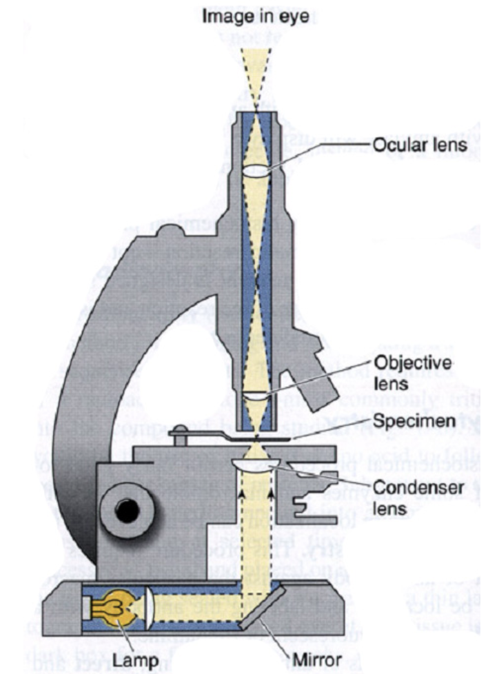
- Illumination Source
- Condenser Lens
- Specimen Stage
- Objective Lens (4x, 10x, 40x to 100x)
- Projection (Ocular) Lens
- Observer
Yields a 2D image capable of a resolution. This is not improvable, as it’s half of the wavelength of visible light, which is .
is the smallest distance between two points on a specimen that can still be distinguished as two separate entities.
Tissue Preparation for Light Microscopy
Cellular features are stained differentially based primarily upon chemical properties.
- Stabilize cellular structure by chemical fixation. (Making it as consistently stable as it would be in the body.)
- Dehydrate and infiltrate tissues with paraffin or plastic. (The presence of water would permit the enzymes to digest and degrade the tissue.)
- Embed fixed tissues in paraffin or plastic blocks. (To permit the cutting.)
- Cut into thin slices of (micrometer) thick; collect sections on slides. (To allow the light to pass through. This is a limitation of the light microscope.)
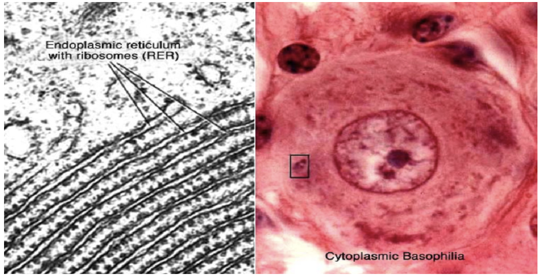
- Re-hydrate and stain with Hematoxylin (a basic dye): Stains acidic structures (e.g. nucleic acids) blue/purple. What do you expect to be stained by Hematoxylin?
- Counter-stain with Eosin (an acidic dye): Stains basophilic structures (e.g. proteins, membranes) red/pink.
H&E staining is routine, but additional dyes and staining techniques may be used to visualize other structures.
Electron Microscopy
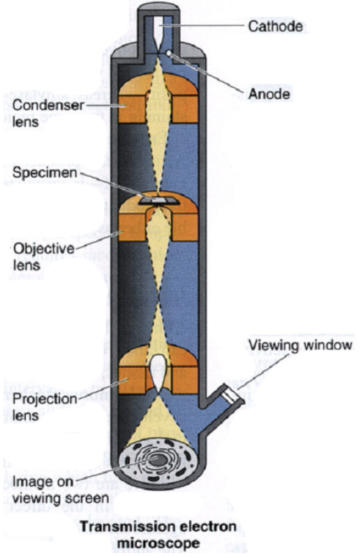
- Illumination Source (Generates electron beam.)
- Condenser Lens
- Specimen Stage
- Objective Lens
- Projection Lens
- View Screen
- Viewing Window & Observer
Yields a 2D image capable of a resolution.
Cellular features are stained with electron-dense, heavy metal stains yielding only a B&W image.
Tissue Preparation for Electron Microscopy
- Tissues are fixed with glutaraldehyde (cross-links proteins) and osmium tetraoxide (cross-links lipids); OsO4 is also an electron-dense “stain”
- Dehydrate and infiltrate tissues w/o plastic.
- Embed and block fixed tissues in plastic. 4. Cut into ultra-thin slices (50 nanometers thick); collect sections on slides.
- Stain sections with heavy metal salts that bind nucleic acids & proteins.
- Visualize in TEM; heavy metal “stains” block electrons to create contrast.
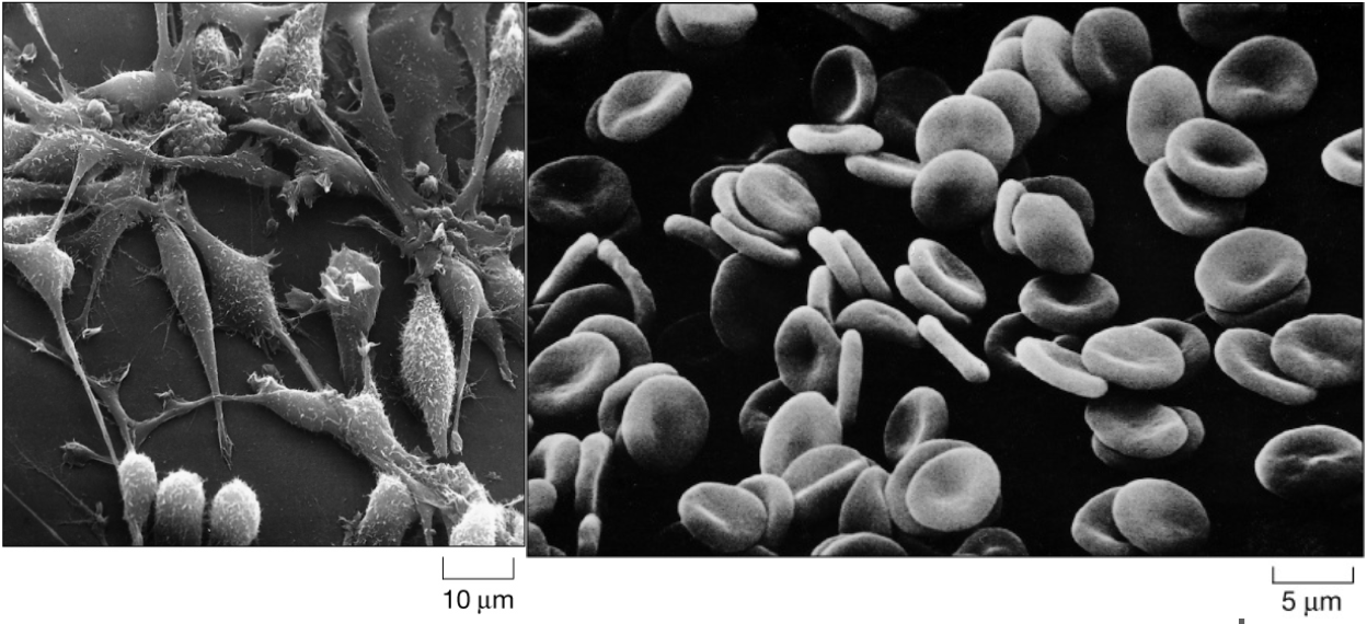
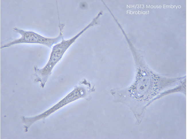
Fibroblasts on the left, RBCs on the right.
Immunofluorescence microscopy
Antigen/antibody reaction, where the antibodies are tagged with a fluorescence dye and the antigen/antibody complex is visualized using a fluorescence microscope.
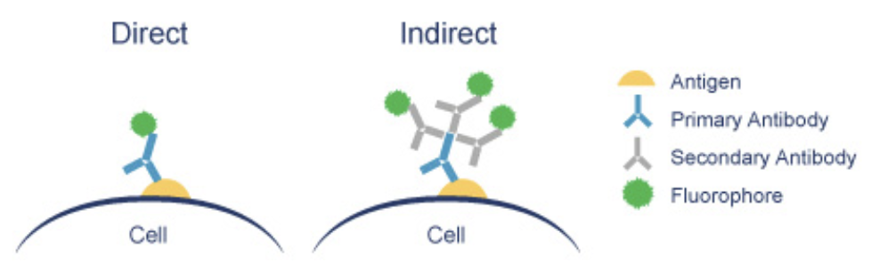
If i’m studying a protein involved in a specific disease, i can study it by using a an antibody which is conjugated with a fluorophore.
Confocal Microscopy (3D Fluorescence)
Resolution: .
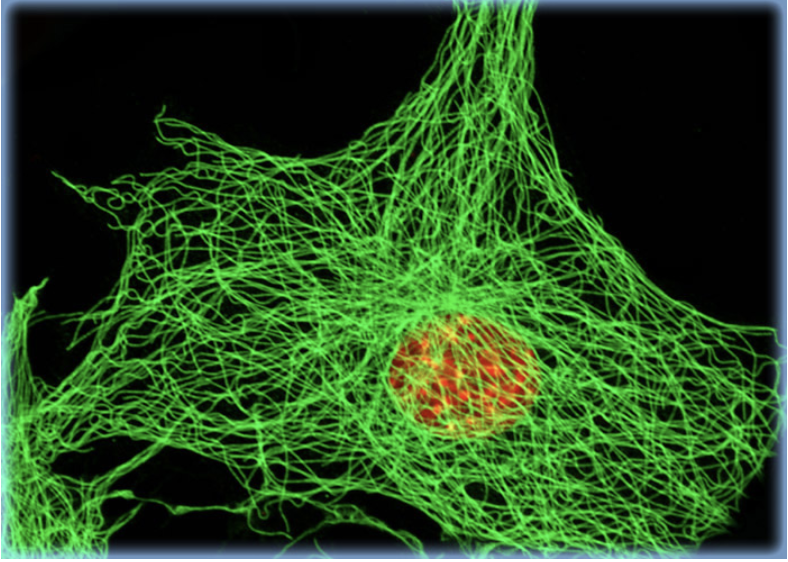
This is a cytoskeleton.
We can see the single filaments of actin.
Green is GFP.
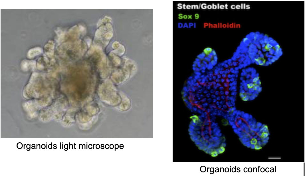
This is a small intestine organoid.
Organoid: growth in vitro of an organ.
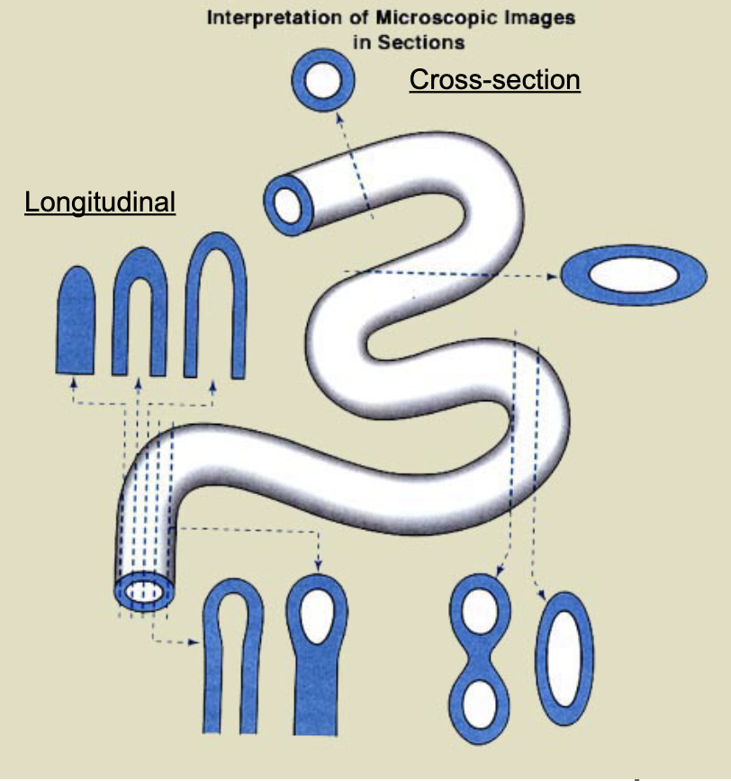
General Issues of Tissue Preparation
Cutting
We can cut the sample in different ways:
- Cross / Transverse Section:
- cutting at a right angle to a specified axis or plane.
- Longitudinal horizontal Section:
- follows the long axis (longitudinal) and divides the object into upper and lower parts (horizontal).
- Longitudinal vertical / Sagittal Section:
- the sagittal plane is parallel to to the body’s midline and divides the sample into two symmetrical sides, on the two sides of the plane.
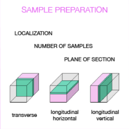
This is very hard but important to classify when recognizing a specimen.
The challenge is being able to view 3D Structures in 2D reliably.
Types of Staining
| Name | Picture | Use |
|---|---|---|
| H&E (Haematoxylin and Eosin Staining) | 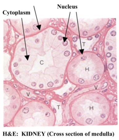 | Haematoxylin binds to basophilic substances (DNA, negatively charged) and stains nuclei in blue-violet. Eosin binds to acidophilic substances (most proteins, positively charged) and stains cytoplasm in red or pink. |
| Picro-Sirius Red Stain | 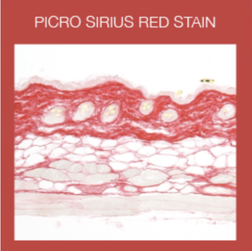 | The Picro-Sirius Red is used in the histological visualization of collagen I and III fibers in paraffin-embedded tissue sections. |
| Red Oil | 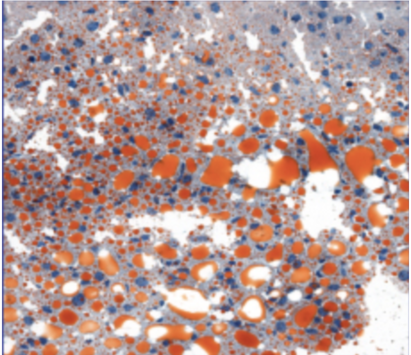 | ORO is used to demonstrate the presence of fat or lipids in fresh frozen tissue sections. It is performed on fresh frozen sections because fixatives containing alcohols, or routine tissue processing, will remove lipids. |
| Alcian Blue / PAS Stain | 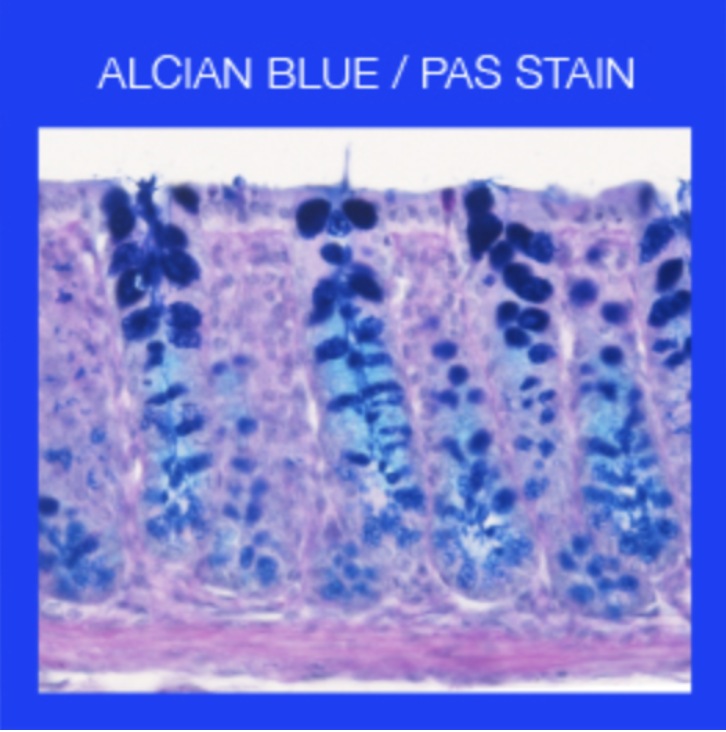 | Alcian Blue stains strongly acidic mucins blue, nuclei pink to red, and cytoplasm pale pink. Periodic acid–Schiff (PAS) detects polysaccharides such as glycogen, glycoproteins, glycolipids, proteoglycans as well as neutral and acidic mucins, allowing for example the identification of hepatic glycogen, intestinal goblet cells and basal laminae. |
LEZIONE 1: Ci fermiamo prima di Golgi’s Black Reaction
Golgi’s Black Reaction & Dispute (accorcia)
Golgi’s “black reaction” (reazione nera) was based on the fixation of nervous tissue blocks in potassium dichromate (2–2.5%) for a variable number of days or weeks (from 1 to 50 days or even longer), followed by immersion in silver nitrate which led to the precipitation of silver chromate fully impregnating cells in the nervous tissue.
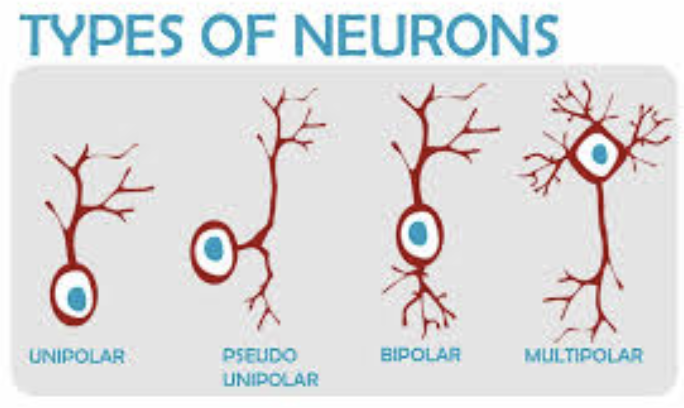
The dispute was a fertile controversy between Camillo Golgi and Ramon y Cajal about the structure of the nervous system.
The nervous system was still considered as a network formed by anastomosing nerve cells, in analogy to the vascular network- the reticular theory of nerve cells continuity: a “reticulum” of interconnected nerve fibers, rather than individual, discrete cells.
Is the nervous system a syncytium (reticular theory) or is it composed of single neurons?
Cajal, paladin of the neuron doctrine, which stated that neurons are individual elements representing the structural and functional units of the nervous system.
Both won the Nobel Prize in 1906.
The metallic impregnation invented by Camillo Golgi in 1873 has allowed the visualization of individual neurons in their entirety, leading to a breakthrough in the knowledge on the structure of the nervous system.
The same experimental data can lead to radically different interpretations depending on theoretical assumptions.
They clashed because of a technical limit of light microscopy: Golgi and Cajal worked with optical microscopes, resolution limit of about 200 nanometers (0,2 um).
Synapses are extremely small (≈20–40 nm gaps between neurons).
Under a light microscope, these gaps cannot be resolved.
Separate neurons therefore appear to touch or fuse together.
The decisive evidence came later:
The debate lasted decades because no available technology could directly show synapses.
The issue was finally resolved with electron microscopy (1950s) → resolution of 0.2 nm revealed:
- a synaptic cleft between neurons
- vesicles releasing neurotransmitters
- membranes remaining separate
Golgi was wrong, Cajal was right. No reticular system, our NS is made of individual neurons.
Tissues
Tissues are functionally related groups of cells that work together.
Types of tissues
- Epithelial – lining and covering or secrete (external / internal)
- Connective – support
- Muscle – movement
- Nervous – control
Structure of tissues
Tissues are made by two elements:
- Cells
- Extracellular matrix
Main differences come from the ratio of cells / ECM.
ECM
Extracellular Matrix (ECM)’s actions:
- Modify the morphology and functions of cells
- Modulate survival of cells
- Regulate the migration of cells
- Direct mitotic activity
- Form junctional associations with cells (via integrins)
The ECM of connective tissue proper is composed of a gel-like ground substance with several fibers embedded.
Cells
Usually classified by:
- Size
- Shape
- Function
- Chemical products
Classification by size
- Lymphocytes about 5-6 µm
- Oocyte 150 µm
- Neurons about mm
- Muscle fiber-syncytium few cm
Classification by shape
Shape and function are correlated
Classification by type of secretion
- Growth factors
- Gas or CO2
Differentiation
The process by which unspecialized cells (stem cells) become specialized to carry out distinct functions.
Stem cells can divide without limits as needed and, under specific conditions, can differentiate into specialized cells.
The first embryonic cells that arise from the division of the zygote are the ultimate stem cells; these stem cells are described as totipotent because they have the potential to differentiate into any of the cells needed to enable an organism to grow and develop.
Blastocyst stage embryo transfer (BET) leads to pregnancy when transfered in a stimulated endometrium.
The embryonic cells that develop from totipotent stem cells and are precursors to the fundamental tissue layers of the embryo are classified as pluripotent.
A pluripotent stem cell has the potential to differentiate into any type of human tissue but cannot support the full development of an organism.
These cells become slightly more specialized, and are referred to as multipotent cells.
A multipotent stem cell has the potential to differentiate into different types of cells within a given cell lineage or small number of lineages, such as a red blood cell or white blood cell.
| Type | Potency |
|---|---|
| Topipotent | Differentiate to enable an organism to grow. |
| Pluripotent | Differentiate into any type of human tissue. |
| Multipotent | Differentiate into different types of cells within a given cell lineage. |
Embryonic Origin of Tissues
After fertilization the zygote gives rise to rapid mitotic cycles, generating many cells to form the embryo.
The first embryonic cells generated have the ability to differentiate into any type of cell in the body and, as such, are called totipotent, meaning each has the capacity to divide, differentiate, and develop into a new organism.
The zygote, or fertilized egg, is a single cell formed by the fusion of an egg and sperm.
As cell proliferation progresses, three major cell lineages are established within the embryo.
Each of these lineages of embryonic cells forms the distinct germ layers from which all the tissues and organs of the human body eventually form.
Each germ layer is identified by its relative position:
- Ectoderm (ecto- = “outer”),
- Mesoderm (meso- = “middle”),
- and endoderm (endo- = “inner”).
The Differentiation Model
Because all cells in the body, beginning with the fertilized egg, contain the same DNA, how do the different cell types come to be so different?
All cells contain the same full complement of DNA, but each type of cell only “reads” the portions of DNA that are relevant to its own function.
In biology, this is referred to as the unique genetic expression.
In order for a cell to differentiate into its specialized form and function, it manipulates those genes (and thus those proteins) that will be expressed, and not those that will remain silent.
The primary mechanism by which genes are turned “on” or “off” is through:
- transcription factors
- and methilation.
A transcription factor is one of a class of proteins that bind to specific genes on the DNA molecule and either promote or inhibit their transcription.
IPS - Induced Pluripotent SCs
The right combination of transcription factors may drive stem cell reprogramming.
Differentiated cells can be reprogrammed to an embryonic-like state by transfer of nuclear contents into oocytes or by fusion with embryonic stem (ES) cells. Thanks Yamanaka!!
This means that unfertilized eggs and ES cells contain factors that can confer pluripotency to somatic cells.
Pluripotent stem cells can be directly generated from mouse embryonic fibroblasts (MEF) cultures by the addition of only a few defined factors.
Which factors?
- He started with 24 TF (Transcription Factors)
- Adding single TF → no iPS
- All together → yes iPS
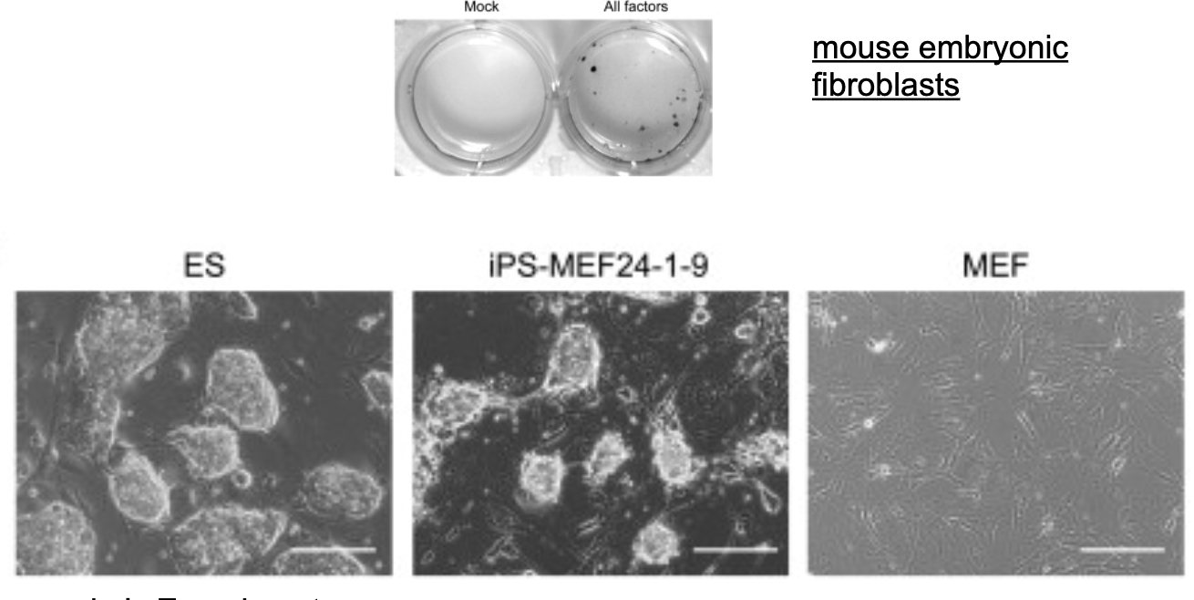
Next, to determine which of the 24 candidates were critical, he examined the effect of withdrawal of individual factors from the pool of 24 candidate genes.
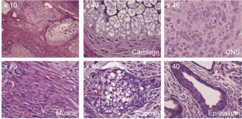
Various tissues present in teratomas derived from iPS-MEF4-7 cells.
Embryonic Derivation
Each of these lineages of embryonic cells forms a distinct germ layers from which all the tissues and organs of the human body eventually form.
| Germ Layer | Gives Rise To: |
|---|---|
| Ectoderm | 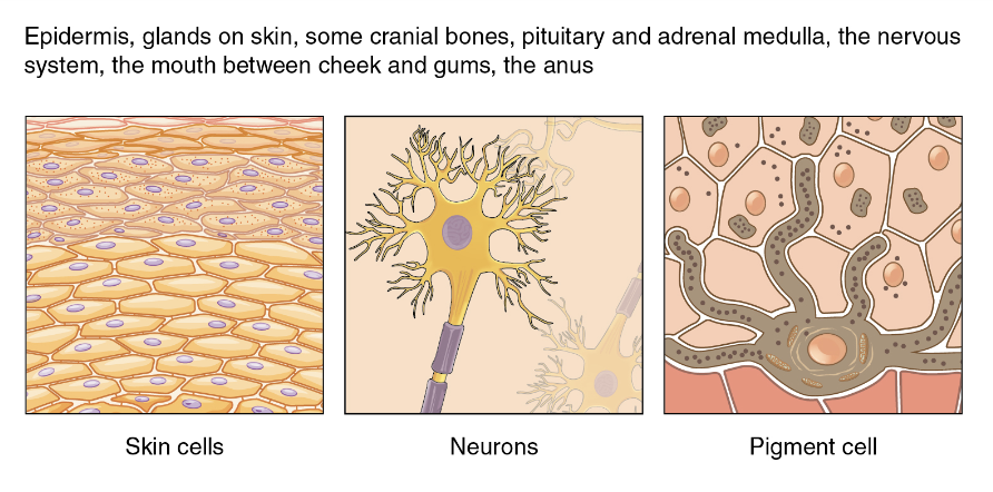 |
| Mesoderm | 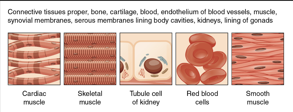 |
| Endoderm | 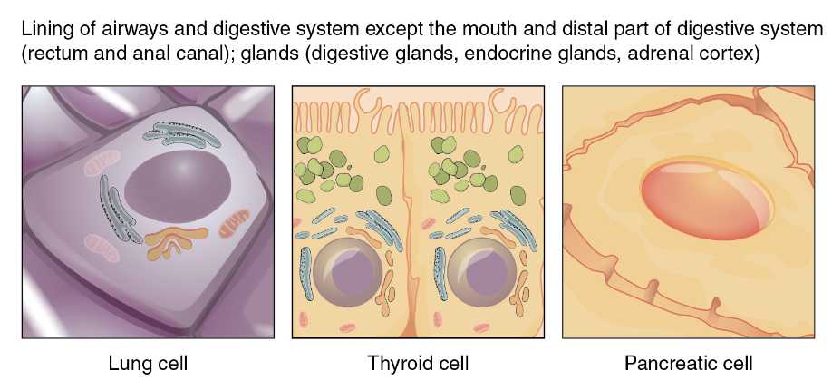 |
Take home message:
- Methods of Microscopy
- Tissue Classification
- Stem Cell/Differentiation
- Embryonic Origin of Tissues
Flashcards
TARGET DECK: MED::I::Morphology and Development::Histology::01 - Introduction
What is the resolution limit of a standard light microscope?
The resolution is , which is approximately half the wavelength of visible light ().
Anki cloze
Hematoxylin is a {1:basic} dye that stains {2:acidic} structures (like nucleic acids) {3:blue/purple}.
Anki cloze
Eosin is an {1:acidic} dye that stains {2:basophilic/protein-rich} structures {3:red/pink}.
What is an artifact in histology?
An unintended result or error that occurs during the collection or preparation of the tissue.
Anki cloze
Electron microscopy yields a 2D image with a resolution of {1:}.
How are specimen prepared for Electron Microscopy to create contrast?
Cellular features are stained with electron-dense, heavy metal stains (like osmium tetraoxide) which block electrons.
Anki cloze
Define the three levels of stem cell potency.
- {1:Totipotent}: Can differentiate into any cell type plus extra-embryonic tissues (new organism).
- {1:Pluripotent}: Can differentiate into any of the three germ layers (all human tissues).
- {1:Multipotent}: Can differentiate into multiple cell types within a specific lineage.
Anki cloze
The three embryonic germ layers are the {1:Ectoderm} (outer), {2:Mesoderm} (middle), and {3:Endoderm} (inner).
What are iPS cells and how are they generated?
Induced Pluripotent Stem cells are differentiated somatic cells reprogrammed to an embryonic-like state by adding specific transcription factors.